KEY FACTS
Terminology
- •
Esophagus atresia (EA) often associated with tracheoesophageal fistula (TEF)
Imaging
- •
Small or absent stomach bubble
- ○
Often difficult to define when stomach is “small”
- ○
Stomach size varies in same fetus over several hours
- ○
Because fetuses breathe amniotic fluid, small amount of fluid may get into stomach if there is TEF
- –
Complete absence suggests no TEF
- –
- ○
- •
Pouch sign
- ○
Transient filling of proximal esophagus with swallowing
- ○
- •
Fetal growth restriction seen in up to 40%
- •
Polyhydramnios rarely develops before 20 weeks
- ○
Fetal swallowing not important part of amniotic fluid dynamics until that time
- ○
- •
Risk for aneuploidy (trisomy 13 and 21)
- •
> 50% have other anomalies
- •
VACTERL (vertebral anomalies, anal atresia, cardiac malformation, TEF/EA, renal anomalies, limb malformations) association in 30%
Scanning Tips
- •
US is poor in detecting EA before onset of polyhydramnios
- ○
Must have high degree of suspicion and perform follow-up scans in setting of small stomach
- ○
- •
Perform focused exam looking specifically at neck and upper chest for esophageal pouch when stomach is small
- ○
Pouch will expand with fetal swallowing
- ○
Distinguish from normal hypopharynx anatomy
- –
Trachea should be easily identified as separate nondistensible structure, relatively thicker wall, and connected to epiglottis
- –
Esophagus located more posterior than trachea
- –
- ○
Determine location of distal end of pouch
- –
Termination in neck worse prognosis than termination in mediastinum
- –
- ○
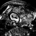
 . No fluid was identified in the stomach throughout gestation, which is suspicious for EA without TEF.
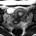
 and was persistently small over multiple scans. The additional finding of polyhydramnios led to a suspicion for EA. A small amount of fluid may get into the stomach if a TEF is present.
 , epiglottis
  , and proximal trachea
  . The esophagus will be just posterior to these structures.


