Esthesioneuroblastoma – delayed postoperative radiosurgery for recurrence at short-term
Skull Base Region
Olfactory Groove
Histopathology
Esthesioneuroblastoma
Prior Surgical Resection
Yes
Pertinent Laboratory Findings
N/A
Case description
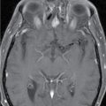
The patient was a 49-year-old male who presented with nasal obstruction and underwent a nasal septal reduction at an outside institution. Postoperatively, he developed progressive hyposmia and left-sided eyelid edema with midface pain. Antibiotic therapy was started without resolution of symptoms, and magnetic resonance imaging (MRI) demonstrated an ethmoid mass eroding through the cribriform plate into the frontal lobe. Transnasal endoscopic biopsy confirmed a Kadish C, Hyams grade 3 esthesioneuroblastoma. The patient underwent a craniofacial resection with positive margins ( Figure 2.5.1 ), followed by external beam radiotherapy (68.4 Gy in 38 fractions) with cisplatin sensitization. Imaging at 6 months revealed recurrence at the margin of radiotherapy field in the bilateral frontal lobes ( Figure 2.5.2 ), for which he underwent stereotactic radiosurgery (SRS) (6 isocenters, 4.9 cm 3 ; 18-Gy margin; 45-Gy maximum dose) ( Figure 2.5.3 ).
Radiosurgery Machine
Gamma Knife – Model C
Radiosurgery Dose (Gy)
18, at 50% isodose line
Number of Fractions
1
Critical Structure
Dose Tolerance
Optic nerve/chiasm
10 Gy maximum point dose
<0.2 cc >8 Gy
Cavernous carotid artery
Tolerant, no evidence of SRS-induced stenosis
Side Effects/Complications
Frequency
Olfactory dysfunction
Usually found on initial diagnosis
Visual dysfunction
<1%
Success Rate/Control Rate
Frequency
Local control
92% at 42 months
Only gold members can continue reading. Log In or Register to continue
Apr 6, 2024 | Posted by drzezo in GENERAL RADIOLOGY | Comments Off on Esthesioneuroblastoma – delayed postoperative radiosurgery for recurrence at short-term
 Stereotactic radiosurgery machines
 Dose tolerances of normal tissues in skull base radiosurgery
 Olfactory groove meningioma – upfront radiosurgery
 Planum sphenoidale meningioma – upfront radiosurgery


