Abstract
Medulloblastoma is the most frequent malignant brain tumor in children. Originating in the cerebellum, they are typically intra-axial tumors. In adults, they represent less than 1% of brain tumors. However, the occurrence of extra-axial medulloblastoma is possible but extremely rare, and slightly more frequent in the adult population. We present a rare case of extra-axial medulloblastoma, diagnosed in a 22-year-old male, through advanced imaging techniques, followed by confirmation through anatomopathological examination.
This case calls attention to the necessity of knowledge of the various diagnostic possibilities when interpreting radiological images, leading to enhanced patient care and furthering our understanding of these exceptional entities.
Introduction
Medulloblastoma is a high-grade malignant tumor classified as an embryonal neuroepithelial tumor [ ]. It represents 15%-20% of central nervous system tumors in individuals under the age of 15 and accounts for one-third of posterior fossa tumors [ ]. Typically, medulloblastomas originate from the cerebellar vermis in childhood, while in adulthood, they arise from the paramedian or lateral regions within the cerebellar hemisphere, where they represent less than 1% of primary brain tumors [ ]. However, a few cases of the posterior fossa extra-axial variant of medulloblastomas have been reported in the literature, most of which are located in the cerebellopontine angle (CPA) [ ]. This raises the issue of differential diagnosis with common tumors of the CPA, such as vestibular schwannomas, meningiomas, and epidermoid inclusion cysts [ ].
We report a case of CPA medulloblastoma in a 22-year-old male who was surgically treated, highlighting that an extra-axial mass in the CPA can indeed be a medulloblastoma.
Case presentation
A 22-year-old male patient with no significant medical history was admitted to the emergency department after experiencing several episodes of vomiting and headaches over the past 2 weeks. During the subsequent neurological assessment, gait disturbances and ataxia were noted over the last 3 weeks. No other neurological disorders were present.
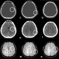
A cranial computed tomography (CT) scan was ordered. The CT scan, performed using a 64-slice GE scanner without contrast administration, revealed a solid, spontaneously hyperdense mass in the left posterior fossa. This mass caused a significant effect on the fourth ventricle, resulting in tri-ventricular obstructive hydrocephalus with periventricular hypodensity consistent with trans-ependymal oedema ( Fig. 1 ).

For further characterization of the lesion, a brain MRI was performed, revealing a well-defined voluminous extra-axial mass in the left cerebellopontine angle (CPA), measuring 37 × 48 × 30 mm. The mass exhibited a hypointense signal on T1-weighted images, isointense signal on FLAIR imaging, and slightly hyperintense signal on T2-weighted images. It was surrounded by a thin layer of cerebrospinal fluid (CSF), confirming its extra-axial origin ( Fig. 2 ).

Diffusion-weighted imaging showed hyperintensity with clear restriction of diffusion (low ADC values). Following the administration of a gadolinium-based contrast agent, the mass exhibited homogeneous enhancement ( Fig. 3 ). Notably, there was pronounced vasogenic edema adjacent to the tumor, evident as a high-intensity signal on FLAIR imaging, and also in the periventricular region ( Fig. 4 ).


Stay updated, free articles. Join our Telegram channel

Full access? Get Clinical Tree


