Abstract
The posterior maxilla is particularly susceptible to bone resorption and poses significant anatomical challenges for surgical interventions, including sinus augmentation and dental implant placement. The Posterior Superior Alveolar Artery (PSAA), which usually courses along the lateral wall of the maxillary sinus, is essential for supplying blood to this area. Nonetheless, variations in the PSAA’s position and diameter can increase the risk of complications during and after surgery. We here report the case of a 36-year-old male patient presented for implant treatment in the posterior maxilla. A Cone Beam Computed Tomography (CBCT) scan revealed a rare anatomical variation of the large PSAA, observed bilaterally on the floor of the maxillary sinus instead of its typical lateral wall position. This unusual finding was considered in preoperative planning to prevent potential complications during implant placement. Preoperative CBCT imaging is essential in identifying uncommon anatomical variations of the PSAA to ensure safe and effective surgical outcomes in the posterior maxilla. Early identification of such variations can guide surgical planning, prevent complications, and enhance patient safety.
Introduction
One of the areas that might undergo alveolar bone atrophy and exhibit poor quality of bone is the posterior maxilla [ ]. All the surgical procedures in the posterior maxilla require a proper recognition of the anatomic details of maxillary sinuses and the possible anatomic variations in this area. Knowledge of anatomical and structural details and the variations of the maxillary sinuses is essential prior to performing any surgery involving the posterior maxilla. The maxillary sinus receives its primary blood supply from the branches of the maxillary artery, namely the posterior superior alveolar artery (PSAA) and the infraorbital artery (IOA). These arteries follow the lateral wall of the maxilla and form an intricate network of anastomoses, both intraosseous and extraosseous, which are crucial for the vascularization of the sinus membrane, periosteum, and lateral wall [ ]. A comprehensive understanding of the anatomy and variations of these arteries is essential to minimize the risk of intra- and postoperative complications during procedures such as sinus augmentation and implant placement in the posterior maxilla. Accidental damage to the PSAA or its anastomoses with the IOA can result in significant bleeding, compromising surgical visibility and performance [ ]. Studies have shown that the location, diameter, and course of the PSAA can vary greatly, influenced by factors such as age, tooth loss, and bone atrophy [ ], necessitating careful preoperative assessment.
To obtain a detailed understanding of the anatomical and functional relationship of the maxillary sinus, 3D imaging is required. Cone Beam Computed Tomography (CBCT) is increasingly recommended for its lower radiation dose and superior ability to accurately delineate the anatomical details of the maxillary sinus, including vascular structures, septa, and other relevant features [ , , ].
Numerous studies have been conducted on the anatomy of the Posterior Superior Alveolar Artery (PSAA), including its course, diameter, and its relationship with adjacent structures. In this study, a case is reported concerning the position and dimensions of the PSAA, which appear to be rare.
Presentation of case
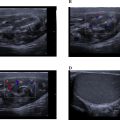
A 36-year-old male patient presented to the Dental School of Shahid Beheshti University of Medical Sciences for implant treatment in the posterior maxillary region. After obtaining initial radiographs, a CBCT scan of the maxilla was performed using NewTom VGi at 110 kVp, automatic mA, voxel size 0.15 mm, and field-of-view of 8 × 12 cm to measure the bone dimensions prior to the placement of dental implants. Incidentally, the posterior superior alveolar artery (PSAA), which is typically visible in the lateral wall of the maxillary sinus, was observed bilaterally in the floor of the maxillary sinus ( Fig. 1 ). The dimensions of the PSAA on the right side were 2.4 × 2.7 mm, and on the left side, they were 1.8 × 2.2 mm ( Fig. 2 ). The distance of the PSAA from the crest of the ridge was measured 15 and 17 mm on the right and left side respectively ( Fig. 3 ). The patient’s CBCT scan report was sent to the periodontics department to ensure that all treatment considerations are taken into account prior to any intervention, such as dental implant placement or sinus lift procedures.


Related posts:
 Breast cancer with medullary features shows a fast and plateau enhancement pattern on magnetic resonance images: A case report
 A rare and life-threatening case of spontaneous hemopneumothorax presenting with severe hemodynamic instability
 Avascular necrosis of lateral femoral condyle in a middle-aged woman from central India
 A very rare case of ileocolic and appendiceal intussusception with acute appendicitis
 Rare case of left epididymo-orchitis complicated by pampiniform plexus thrombosis: A case report
 Hepatic and extra-hepatic hydatid cysts: A case series of radiological and clinical insights
Stay updated, free articles. Join our Telegram channel

Full access? Get Clinical Tree


