fMRI and Wada
Lubdha M. Shah, MD
Key Facts
Wada
Wada test and intraoperative cortical stimulation have been considered gold standards for language representation
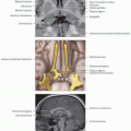
Catheter carotid angiography
Intracarotid administration of sodium amobarbital
Assess consequent cognitive deficit
Disadvantages: Invasive, expensive, nonlocalizing; equivocal results if patient is oversedated and cannot respond
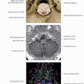
Speech lateralization
Motor hemispheric localization
Memory
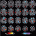
fMRI vs. Wada
Hemispheric language dominance shown to be consistent between the 2 modalities
High correlation between lateralization on fMRI and Wada testing
Many epileptologists now think that fMRI is adequate for language lateralization
In cases where fMRI results are divergent or activation is atypical or absent, Wada may be a helpful presurgical planning tool
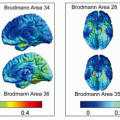
Tasks
Word-generation task significantly more specific than semantic-decision task
Safe
Replicable
Provides more precise anatomic localization within a hemisphere
Memory
Memory task activation does not clearly predict postoperative memory impairment
Therefore, fMRI not a definite substitution for Wada test for overall hemispheric lateralization
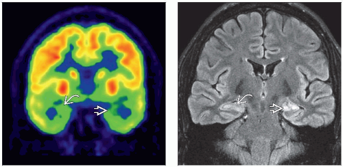 (Left) Coronal PET demonstrates hypometabolism in bilateral mesial temporal lobes in this patient with intractable complex partial seizures. The left temporal lobe
 uptake is 5-10% decreased relative to the right uptake is 5-10% decreased relative to the right  . (Right) Coronal FLAIR MRI shows asymmetric volume loss and hyperintensity in the left mesial temporal lobe . (Right) Coronal FLAIR MRI shows asymmetric volume loss and hyperintensity in the left mesial temporal lobe  in comparison to the right in comparison to the right  . .Related posts:Stay updated, free articles. Join our Telegram channel
Full access? Get Clinical Tree
 Get Clinical Tree app for offline access

|