Foot Motion
Lubdha M. Shah, MD
Key Facts
Imaging Anatomy
Foot/ankle movement results in robust activity within cortex about superior termination of central sulcus in paracentral lobule
Supplementary motor activation is seen
Located in dorsomedial frontal cortex, anterior to leg representation of primary motor cortex
Well-defined somatotopic organization of motor cortex
Motor area devoted to specific region of cortex is proportional to the number of motor units involved in the region’s control
Design
4-minute block design with visual stimulus presentation &/or “on” and “off” commands
Patient asked to dorsiflex and plantar flex each foot at self-paced rate during “on task” and rest during “off task”
Visual cues can be used to control movement rate
Rate can influence activated area of sensory motor cortex
Preprocessing
Distortion, motion, slice timing correction
Coregistration to MPRAGE or T2 anatomic images for overlay
Applications
Presurgical motor mapping for lesions in sensorimotor cortex
Some patients with neurological corticospinal deficits may not be able to carry out foot flexion task
Up to 25% of patients exhibit excessive head motion, rendering the task nondiagnostic
SMA and primary motor cortex for lower extremity may lay adjacent to one another; distinguishing the border between the 2 areas can be problematic
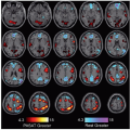
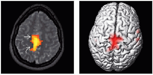 (Left) Axial T2W MRI with overlay of BOLD signal during a bilateral foot flexion task demonstrates activation of bilateral foot sensorimotor regions  . There may be a component of supplementary motor area activation more anteriorly . There may be a component of supplementary motor area activation more anteriorly  . (Right) 3D surface-rendered image in the same patient shows activation in bilateral sensorimotor regions . (Right) 3D surface-rendered image in the same patient shows activation in bilateral sensorimotor regions  related to bilateral foot flexion. related to bilateral foot flexion. |
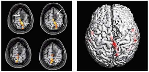 (Left) Axial MPRAGE images with BOLD signal overlay illustrate left sensorimotor activation
 during right foot flexion (top). This area of activation lies posterior to the left superior frontal gyrus lesion during right foot flexion (top). This area of activation lies posterior to the left superior frontal gyrus lesion  . Similarly, left foot flexion (bottom) elicits activation in the right foot sensorimotor area . Similarly, left foot flexion (bottom) elicits activation in the right foot sensorimotor area  . (Right) 3D surface rendering of the same patient with a left superior frontal gyrus oligodendroglioma . (Right) 3D surface rendering of the same patient with a left superior frontal gyrus oligodendroglioma  shows activation in the left foot sensorimotor area shows activation in the left foot sensorimotor area  with right foot flexion. with right foot flexion.Related posts:Stay updated, free articles. Join our Telegram channel
Full access? Get Clinical Tree
 Get Clinical Tree app for offline access

|