KEY FACTS
Terminology
- •
Ganglion cyst: Myxoid tissue encased in fibroblastic collagenous wall with no synovial lining
Imaging
- •
Fluid-filled mass with stalk emanating from joint
- •
Common in wrist but can occur in other joints, including fingers, ankle, knee, hip, and shoulder
- •
Readily diagnosed with ultrasound by characteristic features
- •
Contains hypoechoic fluid
- ○
± septations
- ○
± comet-tail artifacts (due to colloid aggregates)
- ○
- •
Very firm, not compressible
- •
Recent leakage can cause hyperemia and edema of surrounding tissues from inflammatory response
Clinical Issues
- •
Up to 50% of cysts in adults and > 90% in children; wrist ganglia will resolve without treatment
- •
Treated with ultrasound-guided aspiration or surgical excision
- •
Ultrasound-guided aspiration
- ○
Fluid is viscous, larger gauge needle is often required
- ○
Higher success rate alongside flexor tendon sheath, (> 70% vs. 50%)
- ○
Ganglia may become smaller/less symptomatic (partial response)
- ○
Scanning Tips
- •
Locate tell-tale stalk of cyst and trace it back toward joint of origin
- •
Note important relationships of cyst (i.e., to tendons and neurovascular bundle)
- •
Use color Doppler to differentiate from vascular anomaly or nerve sheath tumor, which also appears hypoechoic
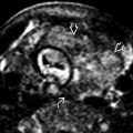
 arising from a defect in the dorsal capsule of the scapholunate ligament
  . The ligament remains functionally intact.
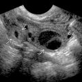
 on the radiovolar aspect of the wrist. The stalk
  of the ganglion extends deeply to lie between the radioscaphocapitate and radiolunotriquetral ligaments
  .
.


