KEY FACTS
Terminology
- •
Bowel herniates through right-sided abdominal wall defect next to normal umbilical cord insertion site
- •
Newborn classification of gastroschisis
- ○
Simple: Isolated, no other bowel anomalies
- ○
Complex: Bowel atresia, necrosis, perforation, volvulus
- ○
- •
Difficult to predict complex gastroschisis in fetus
Imaging
- •
Free-floating bowel, no covering membrane
- •
Bowel dilatation (> 7-10 mm)
- ○
Bowel outside of fetus is often dilated and not predictive of bowel pathology
- ○
Dilated bowel inside fetus more concerning
- ○
> 14-mm bowel diameter most concerning for atresia
- ○
- •
Associations: Growth restriction, oligohydramnios
- •
Can make diagnosis in 1st trimester
- ○
Fetal bowel should be back in abdomen by 12 weeks
- ○
Top Differential Diagnoses
- •
Bowel-only omphalocele
- ○
Bowel not free-floating, covered by membrane
- ○
Cord insertion is not normal: Inserts on omphalocele sac
- ○
- •
Amniotic band syndrome and body stalk anomaly
- ○
Multiple fetal defects
- ○
Clinical Issues
- •
Associated with young maternal age
- ○
Not associated with aneuploidy
- ○
- •
Survival for simple gastroschisis approaches 100%
- •
Complex gastroschisis mortality rates 28-50%
Scanning Tips
- •
Color Doppler shows cord insertion site best
- •
Note appearance of bowel wall
- ○
Thick, echogenic, matted bowel wall is more concerning
- ○
- •
Biophysical profile surveillance later in gestation
- ○
5% risk of intrauterine fetal demise
- ○
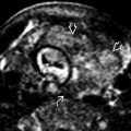
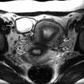
 . The defect is to the right of the umbilical cord insertion site
  .
.
 . Amniotic fluid separates several loops of bowel
  , proving “free-floating” bowel not covered by a membrane. The cord insertion site on the abdominal wall
  is seen well with color Doppler.


