Groove Pancreatitis
Brooke R. Jeffrey, MD
Michael P. Federle, MD, FACR
Key Facts
Terminology
Chronic segmental pancreatitis in pancreaticoduodenal groove
Pancreaticoduodenal groove is defined by head of pancreas (medially) and 2nd portion of duodenum (laterally)
Distal CBD traverses posterior aspect of groove
Imaging
Hypodense nonenhancing mass in groove
Thickened duodenal wall with delayed enhancement
Narrowed lumen ± cysts in wall
Smooth narrowing of terminal CBD and pancreatic duct
Top Differential Diagnoses
Pancreatic carcinoma
Hypodense rounded mass in head; marked atrophy upstream
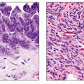
Pathology
Alcohol consumption leads to ↑ viscosity of pancreatic juice → Brunner gland hyperplasia → occlusion of minor papilla
Abnormal minor papilla (absent or rudimentary)
Tumors occluding minor papilla and Santorini duct
Ectopic pancreatic tissue in duodenal wall
Chronic inflammation of terminal CBD, gastric surgery, duodenal/gastric ulcer → fibrosis/scar tissue
Clinical Issues
Postprandial abdominal pain, vomiting
More common in middle-aged men with history of alcohol consumption
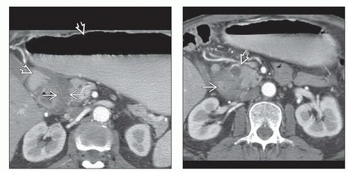 (Left) Axial CECT shows a markedly distended stomach and proximal duodenum  from a gastric outlet obstruction. Note inflammatory exudate in the groove between the 2nd duodenum and the pancreas from a gastric outlet obstruction. Note inflammatory exudate in the groove between the 2nd duodenum and the pancreas  . (Right) More caudal CECT of the same patient shows that the lumen of the 2nd portion of duodenum is markedly narrowed . (Right) More caudal CECT of the same patient shows that the lumen of the 2nd portion of duodenum is markedly narrowed  by inflammation, and there is a small cystic collection in the pancreatic groove by inflammation, and there is a small cystic collection in the pancreatic groove  . . |
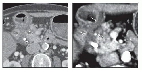 (Left) Axial CECT shows low-attenuation inflammatory exudate in the groove between the head of the pancreas and the 2nd duodenum
 . Note the stent in the common duct . Note the stent in the common duct  placed due to biliary obstruction from the adjacent inflammatory process. (Right) Axial volume-rendered CECT shows that the hypodense inflammation encases the gastroduodenal artery placed due to biliary obstruction from the adjacent inflammatory process. (Right) Axial volume-rendered CECT shows that the hypodense inflammation encases the gastroduodenal artery  and thickens the medial wall of the 2nd duodenum and thickens the medial wall of the 2nd duodenum  . .Related posts:Stay updated, free articles. Join our Telegram channel
Full access? Get Clinical Tree
 Get Clinical Tree app for offline access

|