Gyral/Sulcal Anatomy
Lubdha M. Shah, MD
IMAGING ANATOMY
Lobes
Frontal lobe
Extends to central sulcus
Separated inferiorly & laterally from temporal lobe by sylvian fissure (a.k.a. lateral sulcus)
Parietal lobe
Medially separated from occipital lobe by parietooccipital sulcus
Temporal lobe: Contains auditory cortex
Occipital: Holds visual cortex (i.e., V1, V2, V3)
Insula: Involved in interoception
Covered by lip of cortex: Frontal, parietal, & temporal opercula
Sulci
Frontal lobe
Superior & inferior frontal sulci
Frontal eye field is located at junction of precentral sulcus & caudal-most part of superior frontal sulcus
Precentral, central, postcentral sulci
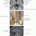
Olfactory sulcus
Contains olfactory bulbs, which transduce & relay odorant information centrally
Orbital sulcus: H-shaped sulcus separating medial, anterior, lateral, & posterior orbital gyri
Parietal
Cingulate sulcus
Surrounds corpus callosum from paraterminal gyrus to isthmus
Marginal branch extends superiorly, lying immediately posterior to central sulcus
Subparietal sulcus is a continuation of cingulate sulcus, separates precuneus from posterior cingulate gyrus
Parietooccipital sulcus marks boundary between cuneus & precuneus as well as parietal & occipital lobes
Intraparietal sulcus separates superior & inferior parietal lobules
Principal functions: Perceptual-motor coordination (for directing eye movements and reaching) & multimodal attention
Temporal
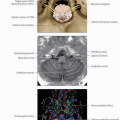
Collateral sulcus
Most mesial temporal sulcus
Lateral limit of parahippocampal gyrus
Superior, middle, inferior temporal sulci
Occipital
Occipitotemporal sulcus separates inferior temporal gyrus, laterally, from occipitotemporal gyrus, mesially
Calcarine separates cuneus from fusiform & lingual gyri
Primary visual cortex along its banks
Lateral occipital sulcus lies on dorsolateral surface
Lunate sulcus in lateral occipital lobe
Transverse occipital sulcus is anterosuperior limit of occipital lobe
Gyri
Frontal
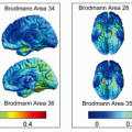
Cingulate gyrus
Anterior cingulate cortex: Processing of salience, pain, reward, emotion, and impulse control
Posterior cingulate cortex: Self-referential cognition, declarative memory, and semantic function
Retrosplenial cortex: Episodic memory and spatial navigation
Gyrus rectus
Orbital gyri
Processes response inhibition and representations of reward, error, emotion, & valuation
Inferior frontal gyrus
Pars orbitalis
Pars triangularis & pars opercularis: Broca area (expressive speech)
Middle frontal gyrus
Part of dorsolateral prefrontal cortex
Executive functioning, working memory, attention
Superior frontal gyrus
Part of premotor cortex; performs initiation & planning motor control
Supplementary motor area
Precentral gyrus: Contains primary motor cortex
Parietal
Postcentral gyrus: Contains somatosensory cortex
Superior parietal lobule
Inferior parietal lobuleRelated posts:

Stay updated, free articles. Join our Telegram channel

Full access? Get Clinical Tree