KEY FACTS
Terminology
- •
Chronic, autoimmune-mediated lymphocytic inflammation of thyroid gland
Imaging
- •
Features vary with stages of disease (acute, chronic, end stage) and extent of involvement (diffuse or focal)
- •
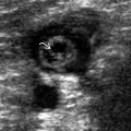
Acute diffuse: Enlarged heterogeneous hypoechoic thyroid with lobulated contour; multiple hypoechoic micronodules throughout with intervening echogenic septa
- •
Acute focal: Discrete, hypoechoic or hyperechoic nodules against normal or altered background thyroid, ± calcifications, halo
- •
End stage: Small, hypoechoic gland with heterogeneous echo pattern
- •
Color Doppler: Vascularity depends on stage and type of involvement
- ○
Acute focal/diffuse: Variable vascularity, focal nodule may mimic benign/malignant thyroid nodule
- ○
- •
Enlarged nodes are common, especially in central neck
Top Differential Diagnoses
- •
Thyroid Non-Hodgkin lymphoma
- •
Graves disease
- •
de Quervain thyroiditis
- •
Riedel thyroiditis (invasive fibrosing thyroiditis)
Clinical Issues
- •
Most common cause of hypothyroidism in USA
- •
Gradual, painless enlargement of thyroid with later atrophy
- •
↑ thyroid peroxidase and antithyroglobulin antibodies
- •
↑ risk of non-Hodgkin lymphoma and papillary carcinoma
Scanning Tips
- •
Be on lookout for developing cancer: Look for nodules that are different from others and for those that are enlarging or contain microcalcifications


