Hemorrhagic Venous Sinus Thrombosis
Shaun R. Rybak
CLINICAL HISTORY
25-year-old female with history of Crohn’s disease presenting with nausea, dehydration, and headache.
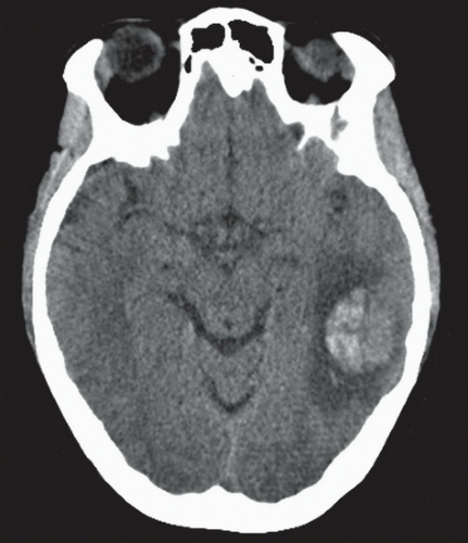 FIGURE 1A |
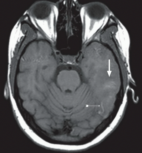 FIGURE 1B |
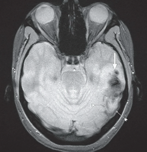 FIGURE 1C |
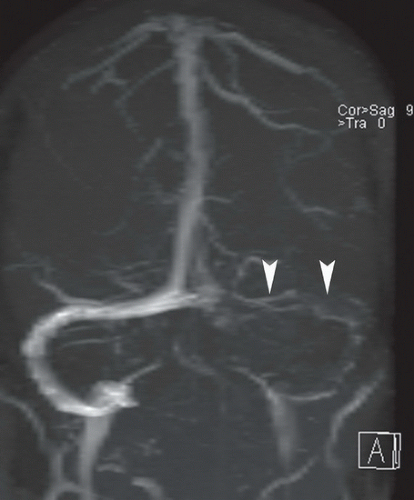 FIGURE 1D |
FINDINGS
Figure 1A: Axial unenhanced computed tomography (CT image) demonstrates a posterior left temporal lobe hemorrhage. Figure 1B: Axial T1 precontrast magnetic resonance imaging (MRI) demonstrates the hematoma (larger arrow) to be of heterogeneous iso- to slightly hyperintense signal relative to the adjacent brain parenchyma. In addition, there is a T1 hyperintense curvilinear cordlike structure posterior to the area of edema in the left temporal lobe (small hatched arrow). Figure 1C: Axial GRE image demonstrates an area of signal dropout with blooming (larger arrow) in the area of the hematoma. There are also thin curvilinear, cordlike structures posterior to the hematoma (small hatched arrows). Figure 1D: Anteroposterior maximum intensity projection (MIP) reconstruction from a magnetic resonance venography (MRV) of the head demonstrates loss of flow-related signal in the left transverse sinus (arrowheads) and sigmoid sinus.
DIFFERENTIAL DIAGNOSIS
Related posts:


Stay updated, free articles. Join our Telegram channel

Full access? Get Clinical Tree


