KEY FACTS
Terminology
- •
Neoplasm of lymphoid tissues in liver
Imaging
- •
No known specific imaging findings for diagnosis of hepatic lymphoma
- •
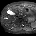
Hepatic lymphoma often favors periportal areas due to high content of lymphatic tissue
- •
Grayscale ultrasound
- ○
Discrete form: Multiple well-defined, hypoechoic masses
- –
Hypoechogenicity due to high cellular density and lack of background stroma
- –
- ○
Infiltrative form: Innumerable subcentimeter hypoechoic foci, miliary in pattern and periportal in location
- –
May be indistinguishable from normal liver
- –
- ○
- •
CECT
- ○
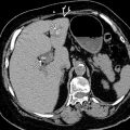
Solid lesions with poor contrast enhancement
- ○
Usually homogeneous density and rarely necrotic
- ○
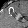
May have thin rim enhancement
- ○
Diffuse, infiltrative, low-density areas
- ○
Top Differential Diagnoses
- •
Metastases
- •
Multifocal/diffuse hepatocellular carcinoma (HCC)
- •
Liver abscesses
- •
Hemangiomas
- •
Focal fat infiltration/sparing
- •
Hepatic cysts
Clinical Issues
- •
Primary hepatic lymphoma is rare
- •
Secondary hepatic involvement is more common
Diagnostic Checklist
- •
Rule out other multiple liver lesions: Metastasis, HCCs, hepatic cysts, abscesses, hemangiomas
- •
Confirmation may require needle biopsy
Scanning Tips
- •
Hepatic lymphoma often has pseudocystic appearance; lack of specular reflection of backwall is helpful clue that lesions are not cystic and rather solid markedly hypoechoic masses