KEY FACTS
Terminology
- •
Accumulation of triglycerides within hepatocytes
Imaging
- •
Diffuse hepatic steatosis
- ○
Increased echogenicity: Liver more echogenic than kidney
- ○
Attenuation of US beam results in poor visualization of diaphragm
- ○
Blurry margins or poorly visualized hepatic and portal veins
- ○
- •
Focal hepatic steatosis
- ○
Geographic hyperechoic area or multiple confluent hyperechoic lesions
- ○
No mass effect with vessels running undisplaced through lesion
- ○
Wedge-shaped/lobar/segmental distribution
- ○
- •
Focal fatty sparing
- ○
Direct drainage of hepatic blood into systemic circulation
- ○
Gallbladder bed: Drained by cystic vein
- ○
Segment 4 or anterior to portal bifurcation: Drained by aberrant gastric vein
- ○
No mass effect with undisplaced vessel
- ○
Top Differential Diagnoses
- •
Steatohepatitis
- •
Fatty cirrhosis
- •
Hemangioma
- •
Metastasis or lymphoma
Clinical Issues
- •
Nonalcoholic fatty liver disease may progress to nonalcoholic steatohepatitis, which may progress to fibrosis and cirrhosis
- •
Cirrhosis is major risk factor for hepatocellular carcinoma
Scanning Tips
- •
Look for undistorted vessels running through regions of steatosis
- •
Ensure overall gain is set correctly; gain set too high can increase apparent echogenicity of liver and mimic steatosis

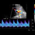
 with a poorly delineated right hepatic vein wall
  and a hardly visible middle hepatic vein
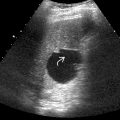
  . Part of the diaphragm is not well visualized due to poor acoustic penetration
  .
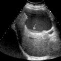
.
Stay updated, free articles. Join our Telegram channel

Full access? Get Clinical Tree


