Hypoxic Ischemic Brain Injury
Shaun R. Wagner
CLINICAL HISTORY
5-year old girl with panhypopituitarism and a recent history of viral illness was found unresponsive by parents, with wet vomitus around her mouth. Paramedics initiated cardiopulmonary resuscitation at the scene.
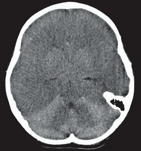 Figure 25A |
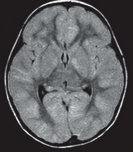 Figure 25B |
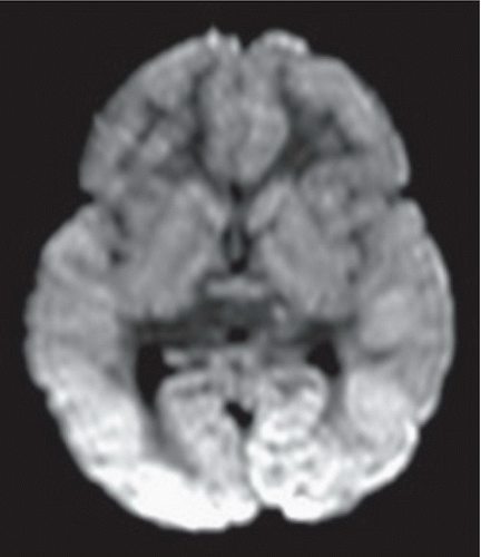 Figure 25C |
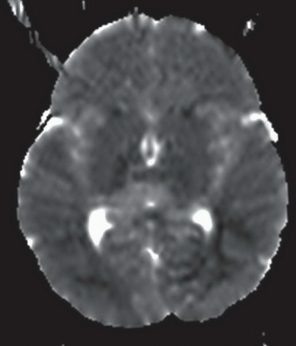 Figure 25D |
FINDINGS
Figure 25A: Axial noncontrast CT images through the brain at the level of the foramen of Monro demonstrates diffuse cerebral edema with loss of the normal gray matter-white matter discrimination. There is relative hyperintensity of the cerebellum juxtaposed against the low-density edematous cerebral hemispheres, the so-called “white cerebellum sign.” Figure 25B: Axial FLAIR image obtained 4 days later at the level of the foramen of Monro demonstrates bilateral hyperintensity of the lentiform nuclei and diffuse cortical hyperintensity, particularly at the occipital lobes. Additionally, there is diffuse thickening of the cortex and effacement of the sulci. Figures 25C and 25D: Axial diffusion-weighted imaging (DWI) (Fig. 25C) and corresponding apparent diffusion coefficient (ADC) map (Fig. 25D) images at the level of the foramen of Monro demonstrate symmetric restricted diffusion throughout the cortex and basal ganglia. The ADC map demonstrates hypointensity in the regions of high signal intensity on DWI, consistent with diffusion restriction.
Related posts:


Stay updated, free articles. Join our Telegram channel

Full access? Get Clinical Tree


