Iliopsoas Neoplasm
R. Brooke Jeffrey, MD
Key Facts
Imaging
Best diagnostic clue
Large retroperitoneal mass secondarily invading psoas
Location
Psoas compartment
Retroperitoneal spaces, including anterior and posterior pararenal spaces
Best imaging tool
CECT, MR
Top Differential Diagnoses
Iliopsoas abscess
Iliopsoas hematoma
Pathology
Etiology
Most often direct invasion by primary retroperitoneal or pelvic tumor
Associated abnormalities
Primary pelvic or retroperitoneal tumor or nodes
Bone destruction of spine
Clinical Issues
Most common signs/symptoms
Back and leg pain
“Psoas” sign with pain on lifting leg
Palpable flank mass
Diagnostic Checklist
Consider well-differentiated liposarcoma if mostly mature fat in lesion
Image interpretation pearls
Myxoid elements in liposarcoma appear cystic on MR
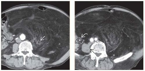 (Left) Axial CECT in a 71-year-old man with a 6 month history of an enlarging left flank mass reveals a large fat-attenuation mass occupying the entire left side of the abdomen and displacing the gastrointestinal tract to the right side of the abdomen. Note the direct infiltration of the left psoas muscle  , most consistent with a liposarcoma. (Right) Axial CECT in the same patient shows the normal right psoas muscle , most consistent with a liposarcoma. (Right) Axial CECT in the same patient shows the normal right psoas muscle  and the normal bowel loops displaced to the right side and the normal bowel loops displaced to the right side  of the abdomen. of the abdomen. |
Related posts:

Stay updated, free articles. Join our Telegram channel

Full access? Get Clinical Tree


