1
Introduction
Lung nodule volumetry is a validated tool to manage indeterminate pulmonary nodules with volume doubling times calculation . While its better reproducibility over uni- or bidimensional electronic caliper–based diameter measurements in assessing nodules sizes is validated , the impact of patient-related technical factors such as inspiratory level on its reliability is debated, with inconsistent results in the literature on the way it could influence its results .
The primary objective of our study was to investigate the effect of inspiratory level on lung nodules volumetry. The secondary objective was to query the amplitude of respiratory effort and nodule-specific characteristics as predictive factors of nodule volume variations.
2
Material and methods
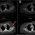
This monocentric retrospective observational study was conducted during the year 2022 in a French university hospital. Patients considered for inclusion underwent unenhanced chest CT including paired acquisitions: an inspiratory CT-scan, immediately followed by an end-expiratory CT-scan, acquired between 2015 and 01–12 and 2021–12–22.
A total of 403 examinations were thus consulted by a single operator (one radiologist with 4 years of training in CT) for preselection and patients were included in our study if they met the following inclusion criteria:
- –
presence of one or more nodule, as defined by the Fleischner Society: Glossary of Terms for Thoracic Imaging
- –
with a greater diameter (on inspiratory CT-scan measured in native axial plane) of at least 6 mm.
Typical and atypical perifissural lung nodules presumed of lymphatic nature, calcified nodules, excavated nodules and nodules issued from exams judged to be technically insufficient/unconform were excluded.
For each included nodule were listed its size (greater diameter on inspiratory CT-scan measured in native axial plane), morphology (solid, ground-glass, part-solid), lobar topography, closeness to the pleura (subpleural or not) and the regularity of its margins (regular/irregular).
CT were acquired on 320-row scanners, with identical technical parameters (collimation : 0,5 <SPAN role=presentation tabIndex=0 id=MathJax-Element-1-Frame class=MathJax style="POSITION: relative" data-mathml='×’>××
×
80 mm , gantry rotation time: 0,275 s , pitch : 0.813, slice thickness/reconstruction increment: 1 mm/0,8 mm ). Raw-image data was worked-off on a mediastinal-window using iterative reconstruction (AID3RD) or deep-learning reconstruction (AiCE).
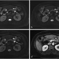
Lung nodule semi-automated segmentation and lung parenchyma segmentation was performed by the same radiologist using Vitrea’s « CT Lung Nodule Analysis » software (version 6.3, Canon Medical Informatics, Minneapolis, Minnesota, USA), with careful manual edition, if judged necessary by the operator.
Finally, nodules and lung volumes variations between inspiratory and expiratory acquisitions were calculated for each pair of exams as followed:
volumevariation(%)=VBMI−VRVBMI×100
Related posts:
 Feasibility of deep learning-reconstructed thin-slice single-breath-hold HASTE for detecting pancreatic lesions: A comparison with two conventional T2-weighted imaging sequences
 Non-visualization of axillary pathological lymph nodes in breast cancer patients on SPECT/CT and during operation
 Development of a deep learning model for the automated detection of green pixels indicative of gout on dual energy CT scan
 Addition of contrast in ultrasound screening for hepatocellular carcinoma
 FDG uptake of pulmonary lesions in synchronous primary lung cancers and lung metastases
 Combination of intrahepatic TARE and extrahepatic TACE to treat HCC patients with extrahepatic artery supply: A case series
Stay updated, free articles. Join our Telegram channel

Full access? Get Clinical Tree


