Abstract
Paget disease of bone is a chronic skeletal disorder characterized by abnormal bone remodeling. This condition, which predominantly affects older adults, can result in a variety of complications, including bone pain, fractures, and deformities. This case report discusses the incidental discovery of monostotic Paget’s disease involving the scapula, leading to brachial plexopathy in a 58-year-old female with a history of colon cancer. The patient had severe bilateral shoulder pain, predominantly on the left, for the past ten years. This pain, exacerbated by overhead activity and limiting daily functions, radiated to the upper limbs without causing weakness or sensory deficits. Clinical examination revealed 4/5 limb power and normal sensation, with significant pain on left shoulder movement and limited elevation. Radiographic investigations, including bilateral shoulder radiographs, contrast-enhanced computed tomography (CT), and cervical magnetic resonance imaging (MRI), identified no displaced fractures but showed diffuse enlargement of left shoulder muscles with fatty infiltration, medullary expansion of the scapula, and reduced left lung volume. MDP skeletal scintigraphy with SPECT/CT confirmed asymmetrical radiotracer uptake and medullary expansion in the left scapula. Further, MRI of both shoulders revealed left-sided expansion, cortical thickening, and fatty replacement of the rotator cuff and deltoid musculature due to denervation pseudohypertrophy from left brachial plexopathy. The right shoulder MRI showed rotator cuff tendinopathy and tears. The findings suggest a diagnosis of monostotic Paget’s disease with secondary brachial plexopathy.
Introduction
Paget’s disease of bone, a chronic disorder of bone remodeling, predominantly affects the elderly [ ]. Although it commonly involves long bones, the pelvis, and the skull, isolated scapular involvement is rare, particularly when associated with brachial plexopathy. This report details an unusual case of monostotic Paget’s disease in the scapula, leading to significant neurological symptoms. Paget’s disease is primarily an osteoclast disease characterized by focal alteration in bone remodeling with resultant atypical osseous structure formation. The growth of bone due to bone remodeling can occur at single site (monostotic) or various sites (polyostotic) [ ]. Paget’s disease was first reported in 1877 in London by Sir James Paget [ ]. Current estimates suggests that approximately 1.5%-8.3% of people suffer from Paget’s disease, making it the second most common metabolic bone disorder globally [ ]. Paget’s disease is rarely encountered before the fourth decade of life. This condition has higher incidence in males. Most patients suffering from Paget’s disease remain asymptomatic [ ]. However, when symptoms do occur, bone pain is the most common complaint of patients. This pain can be dull, constant, boring, and can be deep below the soft tissues [ ]. Bone pain may persist or exacerbate during the night [ ]. It does not spread from one bone to the next and the involvement of new sites are rare. Lesions may continue to progress if the condition is left untreated [ ]. Medical therapy is thought to be the mainstay for the treatment and occasionally surgery can also be performed if complications are severe. However, long-term close follow-up is required to ensure a complete resolution and rule out disease recurrence and possible malignant formation [ ]. Brachial plexopathy is a condition involving compression or injury of the brachial plexus. The brachial plexus is a network of nerves that control movements and sensations in the upper extremity. It is a rare neurological complication associated with Paget’s disease [ ]. Here we present a rare case of monostatic Paget’s disease involving the scapula that induced brachial plexopathy in a patient.
Case presentation
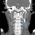
A 58-year-old female presented with a longstanding history of severe bilateral shoulder pain for the last 10 years. Her prior history included colon cancer due to familial adenomatous polyposis (MSH2 mutation), postresection 4 years ago, post-total colectomy with ileoanal anastomosis since 2014, and postradiation therapy. The pain was more pronounced on the left side. The pain was stationary; however, exacerbated by overhead activities, and significantly limits her ability to perform daily activities. The impact of pain on sleep was not reported. The pain also radiated to her neck and upper limbs. There was no history of weakness, sensory deficits, recent trauma, or fever. On physical examination, she had a muscle power of 4/5 in all limbs, normal sensation, and a negative Hoffman’s sign. More pain was noted at the end range of motion in her left shoulder, with limited elevation in both active and passive movements. Bilateral shoulder radiographs revealed no displaced fractures ( Fig. 1 ). Contrast-enhanced CT scans displayed diffuse enlargement of the left shoulder muscles with fatty infiltration and medullary expansion of the scapula, leading to reduced left lung volume ( Fig. 2 ). Cervical MRI excluded any gross disc herniation causing her neuropathy ( Fig. 3 ). MDP skeletal scintigraphy with SPECT/CT confirmed radiotracer uptake, indicating asymmetrical mild medullary expansion of the left scapula with peripheral avidity and deformity in the left first and second ribs ( Fig. 4 ). MRI of both shoulders revealed expansion and cortical thickening with abnormal morphology of the left scapula, and diffuse enlargement with fatty replacement of the left rotator cuff and deltoid musculature due to denervation pseudohypertrophy from left brachial plexopathy, with diffuse intramuscular lipomatosis or congenital disorders such as Klippel-Trenaunay-Weber syndrome ( Fig. 5 ). The right shoulder MRI showed rotator cuff tendinopathy and tears, with a full-thickness tear of the supraspinatus tendon and mild atrophic changes ( Fig. 6 ).


Related posts:
 A rare and life-threatening case of spontaneous hemopneumothorax presenting with severe hemodynamic instability
 A very rare case of ileocolic and appendiceal intussusception with acute appendicitis
 Hepatic and extra-hepatic hydatid cysts: A case series of radiological and clinical insights
 Mid-term follow-up of a fractured flow diverter in the internal carotid artery
 An atypical multiple autoimmune syndrome: A case report including myocarditis
 Persistence of the hypoglossal artery: A rare case report
Stay updated, free articles. Join our Telegram channel

Full access? Get Clinical Tree


