Infected Aortic Stent Graft with Aortitis
Katherine R. Birchard
CLINICAL HISTORY
67-year-old male with chest pain, hypotension, and elevated neutrophil count.
FINDINGS
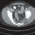
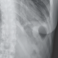
Figure 6A: Serial axial contrast-enhanced CT images of the chest show the aorta with stent graft, and irregular enhancing soft tissue surrounding the arch and descending thoracic aorta (yellow arrows).
DIFFERENTIAL DIAGNOSIS
Infected aortic stent graft with aortitis, acute mediastinal hematoma, small cell carcinoma.
DIAGNOSIS
Infected aortic stent graft with aortitis.
DISCUSSION
Contrast-enhanced computed tomography angiography (CTA) is the first-line imaging modality when acute abnormalities of the aorta are suspected. Findings of infectious aortitis are abnormal periaortic soft tissue with adjacent fat stranding or fluid, and, less commonly, periaortic gas collections. Noncontrast images are also useful, because the periaortic soft tissue enhances with contrast, differentiating it from acute hematoma.1 Infectious aortitis typically occurs in a setting of an atherosclerotic aneurysm (mycotic aneurysm), but can also occur as a result of an infected stent graft, as in this case. Awareness and recognition of imaging findings associated with infected aneurysms are critical for early diagnosis and institution of adequate therapy, in view of the high risk of mortality. IV antibiotic therapy is critical, followed by possible stent removal and revascularization.2,3
Stay updated, free articles. Join our Telegram channel

Full access? Get Clinical Tree


