Abstract
Due to the low frequency and nature of electric injuries, and the different implied mechanisms of trauma, which can manifest in different organs and systems depending on the time of exposure, voltage, amperage and intrinsic resistance of body tissue, the presentation and consequences of the trauma are heterogeneous, and difficult to characterize.
We present the case of a previously healthy adult patient who had a high voltage electric injury, resulting in a diffuse brain leukoencephalopathy with delayed image presentation in MRI as an early complication, and a short review of the mechanisms of electric injury which will help us to understand this pathology and its manifestations in the central nervous system.
Introduction
Electric discharge trauma (EDT) is an infrequent entity, often encountered in young male adults with Laboral exposure. EDT can affect multiple organs and systems, frequently producing local, cardiovascular and neurological injury [ ]. Neurological complications reported [ ] include compromise of central nervous system, peripheral nervous system and even the neuromuscular junction, having diverse presentations such as seizure, intracranial hemorrhage, stroke, hypoxic-ischemic encephalopathy, transverse myelitis and peripheral neuropathy. These complications can appear in the patient with an early or delayed onset after the trauma, and may have variable duration, from temporary to definitive. Most case reports related with neurological EDT complications were diagnosed long time after the moment of the injury [ , ]; which in our case, could be associated in the same episode due to the early onset of symptoms and neuroimaging changes.
Case presentation
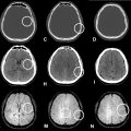
We present the case of a 37 year old male patient, working for an electricity company, who was previously healthy and suffered an electric burn injury after direct contact with a high voltage cable and his right hand. He had 2 episodes of ventricular fibrillation with return to spontaneous circulation after high quality pulmonary resuscitation. Afterwards, he coursed with multiple early epileptic crises, treated with anticonvulsants, which was followed by a diagnostic brain magnetic resonance (BMR), showing no abnormalities at the time given ( Fig. 1 ). The patient received 72 hours of therapeutic hypothermia protocol. Nine days after the initial event, the patient only performed periodical eye opening without contact with the environment. He had flaccid tetraplegia with preservation of brainstem reflexes. In this moment, it was decided to perform a new BMR which showed diffuse and symmetric restricted diffusion of the subcortical, periventricular white matter, involving the corpus callosum and Putamina, with subtle representation in FLAIR sequences; these findings were associated with narrowing of the sulci and subarachnoid spaces ( Fig 2 ).


Stay updated, free articles. Join our Telegram channel

Full access? Get Clinical Tree


