Abstract
Hepatic angiomyolipoma (HAML) is a rare, benign tumor characterized by a mixture of fat, smooth muscle, and blood vessels. We present the case of a 37-year-old male with a history of tuberous sclerosis complex who underwent a CT scan to evaluate abdominal manifestations of the disease. The imaging was performed with and without contrast enhancement and analyzed across arterial, venous, and delayed phases. CT imaging revealed hepatomegaly with a large, ill-defined, nonencapsulated heterogeneous mass composed of both fatty and soft tissue components. Postcontrast enhancement demonstrated a heterogeneous pattern with arterial-phase vascular structures (branches of the hepatic artery) and venous-phase elements (branches of the portal vein network). The lesion extended to the superior, inferior, anterior, and posterior liver borders while preserving the biliary hilum. These imaging findings were consistent with hepatic angiomyolipoma; however, no biopsy was performed for histopathological confirmation. This study aims to highlight the CT characteristics of hepatic angiomyolipoma.
Introduction
Hepatic angiomyolipoma (HAML) is an uncommon benign tumor composed of a combination of fat, smooth muscle, and blood vessels. HAML is extremely rare, with only a few hundred cases reported in the literature [ ]. It predominantly occurs in middle-aged adults, with the mean age at diagnosis typically in the fourth to fifth decade of life. It is more frequently observed in women, with a female-to-male ratio of approximately 3:1. This gender predilection is thought to be linked to hormonal influences.Although angiomyolipomas primarily occur in the kidneys, their hepatic counterpart is increasingly recognized, often discovered incidentally during imaging for unrelated conditions. While most cases are asymptomatic, larger lesions may lead to complications such as hemorrhage or hepatic dysfunction. Diagnosis relies heavily on imaging techniques, particularly computed tomography (CT) and magnetic resonance imaging (MRI), which facilitate accurate characterization and differentiation from other hepatic lesions [ ]. On ultrasound, HAML typically appears hyperechoic, sometimes with mixed echogenicity. On CT, it presents hypodense lesion on noncontrast scans, with heterogeneous arterial enhancement and possible washout in later phases.
On MRI, HAML appears hyperintense on T1-weighted sequences due to its fat content, exhibits variable signal intensity on T2-weighted images, and demonstrates signal loss on fat-suppressed sequences. Postcontrast imaging reveals heterogeneous enhancement. Management strategies range from conservative monitoring to surgical intervention, depending on tumor size and symptomatic presentation. This article aims to provide a comprehensive overview of the clinical presentation, imaging characteristics, and management options for hepatic angiomyolipoma, emphasizing the importance of early recognition and appropriate treatment to prevent complications.
Case presentation
A 37-year-old male patient with a history of tuberous sclerosis complex and childhood seizures presented for a scannographic evaluation of his condition. Clinically, the patient did not report abdominal pain, vomiting, or gastrointestinal disturbances such as constipation or diarrhea. Notably, there was no jaundice, palpable abdominal mass, or any other digestive or extra-digestive symptoms. However, the patient exhibited more pronounced skin lesions on the face, including tubers and angiofibromas. The patient had previously undergone a thorough assessment, including liver and kidney function tests and an ionogram, all of which were normal. An abdominal CT scan with contrast enhancement was performed, with imaging analyzed in the arterial, venous, and delayed phases.
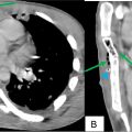
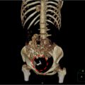
Imaging revealed an enlarged liver (LH = 17 cm), characterized by a heterogeneous appearance, containing a large, ill-defined, non-encapsulated mass composed of fatty and tissue components. The mass demonstrated heterogeneous enhancement after contrast injection, with vascular structures of both arterial (branches of the hepatic artery) and venous (branches of the portal vein network) types. It extended to the superior, inferior, anterior, and posterior borders of the liver, while sparing the biliary hilum.
The mass was localized in segments I, IVa, IVb, VII, and VIII, measuring 123 × 124 × 138.9 mm length × anteroposterior × height). Given the patient’s clinical history, the imaging features were suggestive of a hepatic angiomyolipoma. Correlation with the patient’s clinical and histological data was recommended. The portal and hepatic veins were of normal caliber and patent, with thinintra and extra hepatic biliary pathways.
This scanographic appearance was consistent with hepatic angiomyolipoma. However, the patient did not undergo a biopsy for pathological confirmation. The patient’s condition evolved favorably, and no medical, interventional, or surgical treatment was deemed necessary due to his asymptomatic status and the overall good prognosis ( Figs. 1-4 ).


Related posts:
 Breast cancer with medullary features shows a fast and plateau enhancement pattern on magnetic resonance images: A case report
 A rare and life-threatening case of spontaneous hemopneumothorax presenting with severe hemodynamic instability
 Primary sternal osteomyelitis in an immunocompetent adolescent
 Avascular necrosis of lateral femoral condyle in a middle-aged woman from central India
 A very rare case of ileocolic and appendiceal intussusception with acute appendicitis
 An incidental finding of an ectopic intrauterine device (IUD) in the urinary bladder wall in a female presented with missed abortion
Stay updated, free articles. Join our Telegram channel

Full access? Get Clinical Tree


