Abstract
Objective
Alternative medical imaging techniques are necessary to address the limitations of magnetic resonance imaging (MRI). Therefore, this study aimed to develop an ultrasonographic method that integrates anterior and posterior approaches for measuring the entire length of the anterior cruciate ligament (ACL). We validated this method by identifying the middle ACL during arthroscopy and comparing the results to those of MRI. We hypothesized that the ACL length measurements obtained via ultrasonography and MRI would not differ significantly and that the posterior approach would provide a longer visual field of the ACL than the anterior approach.
Methods
Thirty-six patients (21 males, 15 females) diagnosed with meniscal injury or internal knee derangement were included. During arthroscopy, the surgeon identified the middle ACL using Ti-Cron™ sutures. Ultrasonographic approaches from the anterior and posterior perspectives were used to identify the distal and proximal ACL, respectively. The ACL length was measured using both ultrasonography and MRI, and the visual fields from both approaches were compared.
Results
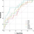
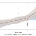
One participant was excluded because of a torn ACL, and seven participants were excluded because of poor ultrasonographic image quality. The ACL length of the 28 included patients did not differ significantly between ultrasonography and MRI, with a moderate correlation between the two measurements. The visualized proportion of the ACL was greater through the posterior approach than through the anterior approach.
Conclusions
This ultrasonographic method visualizes the entire ACL length by combining anterior and posterior approaches, with the posterior offering a more extensive and clinically significant visual field.
Introduction
Anterior cruciate ligament (ACL) rupture is a prevalent issue in sports medicine, with an estimated incidence of 30–78 per 100,000 person-years [ ]. Arthroscopy is the gold standard for diagnosing ACL injuries, whereas magnetic resonance imaging (MRI) is the primary diagnostic imaging modality in contemporary medical practice and research owing to its practicality [ ]. MRI can reveal disrupted fiber continuity in the ACL, often presenting as a thickening or wavy pattern in cases of ACL injury [ ]. The diagnostic sensitivity and specificity of MRI in detecting ACL tears are estimated to be approximately 87% and 93%, respectively [ ]. Currently, MRI is the gold standard for ACL imaging because it provides detailed visualization of the ligament. However, clinical researchers have not abandoned the use of alternative medical imaging tools, such as ultrasound, to evaluate ACL injuries.
Ultrasonography has been used to diagnose ACL injuries by identifying indirect indicators, such as the hypoechoic intercondylar notch sign in the posterior transverse view, posterior capsule protrusion, a wavelike posterior cruciate ligament (PCL) in the posterior longitudinal view, and the morphology of the distal ACL in the anterior longitudinal view [ ]. Another type of assessment involves ultrasonography-based anterior drawer tests, which focus on proximal tibial anterior translation induced by gravity or manual compression using a posterior approach [ , ]. Ultrasonography has 86.8%–100% sensitivity and 87.5%–97.7% specificity for diagnosing acute ACL injuries, but it primarily visualizes indirect signs of ACL injuries or the distal portion of the ACL. Direct observation of an injured ACL primarily focuses on visualizing the distal portion of the ACL using the anterior approach. However, clinical observations have indicated that most ACL injuries occur in the proximal to mid-substance area [ ]. Inadequate visualization of the ACL compromises the efficacy of ultrasonography, particularly in patients with chronic injuries. Surgeons who are proficient in implementing ultrasonographic methods can simultaneously validate new methods for visualizing the proximal ACL via ultrasonography during arthroscopy.
The proficiency of ultrasound-guided aspiration of proximal ACL cysts has been established; notably, previous studies used a posterior approach for cysts rather than for the ACL [ , ]. However, these findings underscore the potential of visualizing the proximal ACL and highlight the need for further validation. The anterior and posterior approaches to ultrasonography provide distinct perspectives on ACL visualization. Integrating the distal and proximal ACLs in the anterior and posterior approaches, respectively, during ultrasonography allows for a comprehensive view of the entire ACL length. Validation of our method using arthroscopy and MRI is essential to assess its accuracy and evaluate its usefulness.
In this study, we aimed to visualize the entire ACL length using anterior and posterior sonographic approaches. We applied an arthroscopic method to validate the sonographic findings, delineated the ACL border using surgical sutures, and further validated our results using MRI. We hypothesized that there would be no significant difference in the length of the ACL as measured by ultrasonography and MRI and that a correlation would exist between these two imaging tools. Furthermore, we calculated the proportion of visible ACL in the anterior and posterior approaches. We speculated that the percentage of the visualized ACL length would be greater using the posterior approach than using the anterior approach.
Materials and methods
Study design and population
This cross-sectional, observational study was approved by the Institutional Review Board of our center and complied with the ethical standards enshrined in the 1964 Declaration of Helsinki (2013 amendment). Patients were recruited from the Department of Orthopedic Surgery and received an explanation regarding the study purpose and procedures. The patients were free to choose whether to participate in this study, and 36 patients provided written informed consent to participate and for the publication of their innominate data.
The inclusion criteria were as follows: age >18 years, meniscal injury or internal knee joint derangement diagnosed on the basis of clinical manifestations and MRI, indications for arthroscopic surgery, and consent to perform ACL measurement intraoperatively using ultrasonography. The exclusion criteria were as follows: a history of ipsilateral knee surgery in the past 2 years, ACL laxity determined by probing during arthroscopy, and unclear ACL boundaries or surgical sutures during ultrasonography leading to measurement failure.
All patients underwent arthroscopic knee surgery. After anesthesia, the patients were positioned supine, and the anteromedial and anterolateral portals were established. The ACL was explored after meniscal repair or other required procedure. The resistance force of the ACL was initially verified by probing. Patients with ACL laxity during surgery were excluded from the study. Subsequently, a number 5 Ti-Cron TM polyester suture (Medtronic, Dublin, Ireland) was folded, and the loop end was introduced into the knee joint via the anteromedial portal. The suture was wrapped around the midpoint of the ACL to serve as a landmark. The anterior and posterior boundaries of the ACL were delineated using Ti-Cron TM sutures ( Fig. 1 ). The free end of the suture was left exposed outside the skin to facilitate easy access to the ultrasonographic dynamic test to verify the ACL location. We then used the anterior and posterior ultrasonographic approaches to detect the ACL.

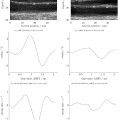
In the anterior approach, the knee joint is flexed at 90° as previously described [ , ]. A SuperSonic Ultrasound system with a curvilinear 1–6-MHz transducer (Supersonic Imagine, Aix-en-Provence, France) with a wide view was required to capture both the distal femur and the proximal tibia to visualize the ACL between them. The image depth was set to 5–6 cm. The transducer was positioned at the midline of the patellar tendon, and its long axis was initially aligned parallel to the patellar tendon. Images acquired from this position showed the patella, patellar tendon, and tibial plateau, with a hyperechoic line almost perpendicular to the patellar tendon. Then, the transducer was externally rotated by ∼10° to obtain a longitudinal view of the distal ACL. The distal ACL appeared as a hypoechoic, band-like structure attached to the tibial plateau, with a visible hyperechoic Ti-Cron TM suture ( Fig. 2 and Video 1 ; the video files are provided in the supplementary materials). The distance between the Ti-Cron TM suture and the ACL tibial insertion (L tibia-suture ) was measured, and the length of the visible ACL in the anterior approach was determined. These measurements were performed along the posterior edge of the ACL.

The knee remained straight in the posterior approach, with a pillow positioned under the ankle or lower leg to raise the knee joint off the table. The transducer for longitudinal views of the ACL was placed around the popliteal fossa, deviating slightly to the medial aspect. The image depth varied between 6 and 8 cm, with the long axis of the transducer aligned approximately parallel to the lower limb, and the proximal end of the transducer was rotated slightly toward the lateral side. The medial aspect of the lateral femoral condyle was identified; next, the transducer was slid medially and tilted gradually to locate the hyperechoic slope on the roof of the femoral intercondylar notch (i.e., the Blumensaat line) on plain films. The hypoechoic ACL was visualized adjacent to this slope, extending distally toward the tibia. The PCL at the tibial end was also visible in the same visual field, ensuring a straight alignment of the ACLs ( Fig. 3 and Video 2 ; the video files are provided in the supplementary materials). The distances between the Ti-Cron TM suture and the uppermost point of the ACL (L femur-suture ) were measured, and the lengths of the visible ACL in the posterior view were recorded ( Fig. 4 ). The aforementioned measurements were performed along the posterior edge of the ACL. The entire ACL length was defined as follows:
ACLlength=Ltibia−suture+Lfemur−suture


The percentage of visible ACL from the anterior and posterior approaches was calculated as follows:
Visiblepercentagefromtheanteriorapproach=(VisualizedACLinanteriorview)/(Ltibia−suture+Lfemur−suture).
Related posts:
 Quantitative Analysis of Contrast-Enhanced Ultrasound Images of Brain-Dead Donor Livers: Prediction of Early Allograft Dysfunction
 Safety of Shear Wave Elastography as Evidenced From Carotid Artery Strain and Strain Rate Induced by Acoustic Radiation Force Impulse and Arterial Pulsations
 Ultrasound Shear Wave Elastography and Shear Wave Dispersion: Correlation with Histopathological Changes in Autoimmune Hepatitis Patients
 Diagnosis of Salivary Gland Tumors Using Ultrasound Radiomics
 Empirical Model of Focused Ultrasound-Mediated Treatment for Chemotherapy Delivery to Brain Tumors
 High-Frequency Ultrasound Characterization of Periodontal Soft Tissues Pre- and Post-Bacterial Inoculation
Stay updated, free articles. Join our Telegram channel

Full access? Get Clinical Tree


