Jumped Facets
Christopher J. Karakasis
CLINICAL HISTORY
28-year-old patient with low back pain and lower extremity weakness following a motor vehicle collision.
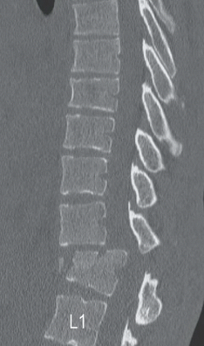 FIGURE 9A |
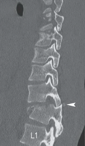 FIGURE 9B |
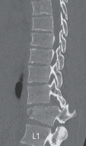 FIGURE 9C |
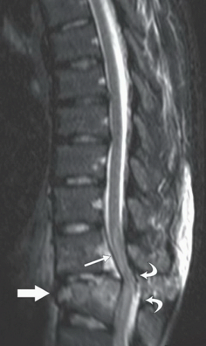 FIGURE 9D |
FINDINGS
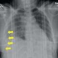
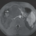
Figure 9A: Sagittal CT of the thoracic spine at midline demonstrates anterior compression of T12 with displaced anterior fracture fragments and anterolisthesis of the T11 vertebral body relative to T12. There is resultant severe spinal canal stenosis. Figures 9B and 9C: Sagittal CT of thoracic spine to the left (Fig. 9B) and right of midline (Fig. 9C) demonstrate bilateral jumped facets, with a small displaced fracture of the left T12 superior articular process (arrowhead in Fig. 9B). Figure 9D: Sagittal STIR MR image demonstrates high signal intensity bone marrow edema throughout the T12 vertebral body caused by fracture. There is separation of the anterior longitudinal ligament (thick arrow) from the anterior aspect of T12, and lifting of the posterior longitudinal ligament (thin arrow) from the posterior T11 body with acute hemorrhage underlying the ligaments. There is widening of the interspinous space with signal abnormality within the interspinous and supraspinous ligaments as well as discontinuity of ligamentum flavum (curved arrows), indicating posterior ligamentous complex injuries. There is severe spinal stenosis and increased signal of the spinal cord as a result of acute compressive cord injury.
Related posts:


Stay updated, free articles. Join our Telegram channel

Full access? Get Clinical Tree


