Limbic System
Karen L. Salzman, MD
Terminology
Definitions
Limbic lobe
Phylogenetically older cortex
Fewer layers than neocortex
Major role in memory, olfaction, emotion
Composed of subcallosal, cingulate, parahippocampal gyri + hippocampus, dentate gyrus, subiculum, entorhinal cortex
Limbic system
Limbic lobe
Plus some subcortical structures (e.g., amygdala, mammillary bodies, septal nuclei, etc.)
Gross Anatomy
Overview
Limbic lobe formed by nested “C-shaped” arches of tissues surrounding diencephalon, basal ganglia
Outer arch
Largest of the three arches
Extends from temporal to frontal lobes, comprised of
Uncus (anterior end of parahippocampal gyrus)
Parahippocampal gyrus (swings medially at posterior temporal lobe, becomes isthmus of cingulate gyrus)
Cingulate gyrus (anterosuperior continuation of parahippocampal gyrus)
Subcallosal (paraolfactory area) is anteroinferior continuation of cingulate gyrus
Curves above callosal sulcus (continuous with hippocampal sulcus of temporal lobe)
Middle arch
Extends from temporal to frontal lobes, comprised of
Hippocampus proper (Ammon horn)
Dentate gyrus
Supracallosal gyrus (indusium griseum, a thin strip of gray matter that extends from dentate/hippocampus all the way around corpus callosum to paraterminal gyrus)
Paraterminal gyrus (below corpus callosum rostrum)
Curves over corpus callosum, below callosal sulcus
Inner arch
Smallest arch
Extends from temporal lobe to mamillary bodies
Comprised of fornix, fimbria
Imaging Anatomy
Overview
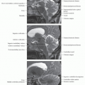
Hippocampus
Curved structure on medial aspect of temporal lobe that bulges into floor of temporal horn
Consists of two interlocking “U-shaped” gray matter structures
Hippocampus proper (Ammon horn) forms more superolateral, upside-down U
Dentate gyrus forms inferomedial U
Has three anatomic subdivisions
Head (pes hippocampus): Most anterior part, oriented transversely; has 3-4 digitations on superior surface
Body: Cylindrical, oriented parasagittally
Tail: Most posterior portion; narrows then curves around splenium to form indusium griseum above corpus callosum (CC)
Ammon horn (hippocampus proper)
Subdivided into four zones (based on histology of main cell layers)
CA1 (Sommer sector): Small pyramidal cells (most vulnerable; commonly affected by anoxia, mesial temporal sclerosis)
CA2: Narrow, dense band of large pyramidal cells (“resistant sector”)
CA3: Wide loose band of large pyramidal cells
CA4 (end-folium): Loosely structured inner zone, enveloped by dentate gyrus
Blends laterally into subiculum
Subiculum forms transition to neocortex of parahippocampal gyrus (entorhinal cortex)
Covered by layer of efferent fibers, the alveus
Alveus borders temporal horn of lateral ventricle ventricle
Forms fimbria → crus of fornix
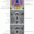
Fornix
Primary efferent system from hippocampus
Four parts
Crura (arch under CC splenium, form part of medial wall of lateral ventricles)
Commissure (connects crura)
Body (formed by convergence of crura, attached to inferior surface of septum pellucidum)
Columns (curve inferiorly to mammillary bodies, anterior thalamus, mamillary bodies, septal nuclei)
AmygdalaRelated posts:

Stay updated, free articles. Join our Telegram channel

Full access? Get Clinical Tree