KEY FACTS
Terminology
- •
Placenta previa (PP) : Placenta covers cervix internal os (IO)
- •
Low-lying placenta (LLP) : Placenta edge < 2 cm from IO
- •
Normal: Placenta edge 2 cm or more from IO
- •
Avoid “marginal” or “partial” PP (deemed confusing)
Imaging
- •
Placental tissue near cervix is hallmark finding
- ○
Sagittal view is best
- ○
- •
Transvaginal US (TVUS) is essential for diagnosis
- ○
Best way to evaluate lower uterine segment (LUS)
- ○
Measure distance between placental edge and IO
- ○
Marginal placental vessels count as placenta
- ○
- •
Important associations
- ○
Morbidly adherent placenta (placenta accreta spectrum)
- –
↑ risk if prior cesarean section (C-section)
- –
Greater risk if multiple prior C-sections
- –
- ○
Vasa previa: Fetal vessels cross IO
- ○
Top Differential Diagnoses
- •
Overdistended maternal bladder: Pushes front and back of uterus together; mimics long cervix and low placenta
- •
Focal myometrial contraction: Shortens uterus, “pulls” placenta low
- •
Placental abruption: Isoechoic blood clot mimics placenta
Clinical Issues
- •
LLP or PP is seen in 2% of all midgestation studies
- •
> 90% will resolve: Follow-up at 32 and maybe 36 weeks
- •
C-section for PP, LLPs might deliver vaginally
Scanning Tips
- •
Scan whole uterus before determining placenta location
- •
TVUS if any placenta seen in LUS
- •
Use color Doppler: Find cord insertion, rule out vasa previa
- •
Move baby head out of pelvis
- ○
Put bed in Trendelenburg (head lower than feet)
- ○
2nd person in room to push baby head up
- ○
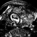
 is several centimeters over the internal cervical os
  . It is not surprising that this case of asymptomatic placenta previa (PP) did not resolve with advancing pregnancy.
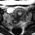
 close to, but not covering, the cervical os
  . There is a 90% chance this will resolve with advancing pregnancy. Follow-up at 32 weeks was recommended.
Stay updated, free articles. Join our Telegram channel

Full access? Get Clinical Tree


