Abstract
Giant arachnoid granulations have been previously reported in the literature, with the largest documented measuring 6 cm and usually associated with significant clinical symptoms. Herein, we present a patient with a massive, irregular-shaped arachnoid granulation occupying both transverse sinuses, a finding that has not yet been reported. The patient, who had a history of transvenous CSF-venous fistula (CVF) embolization, presented with persistent left occipital headaches. Digital subtraction myelography (DSM) to investigate a residual or recurrent CVF yielded negative results. Contrast enhanced brain MRI showed a nonenhancing lobulated lesion in the expected location of, but distinct from the transverse sinuses. The patient was later started on indomethacin, which completely resolved his headaches, consistent with the diagnosis of hemicrania continua. This indicated that the arachnoid granulation was an incidental finding despite its unusual size.
Introduction
Arachnoid granulations are outgrowths of subarachnoid membranes in the dural sinuses in which CSF flows to the venous system [ ]. They constitute the major route of resorption of CSF fluid [ ] and are known to be responsible for intracranial volume regulation [ ]. They are macroscopically visible to the naked eye and increase in size and number with age, in response to an increase in CSF pressure in the subarachnoid space [ , ]. Giant arachnoid granulations (GAGs) have been increasingly reported in the literature, with sizes ranging up to 6 cm. They can be asymptomatic, or present with heterogenous signs and symptoms, with headaches being the most common presentation [ ]. In this report, we describe the diagnosis of an extremely large arachnoid granulation in a 45-year-old male who underwent diagnostic workup with MRI and lateral decubitus DSM at our institution.
Case report
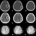
A 45-year-old male presented with persistent left occipital headaches. He had undergone transvenous embolization of a spinal CSF-venous fistula (CVF) in the past. He underwent digital subtraction myelography with delayed CT myelogram (CTM) in our institution to investigate a possible residual or recurrent CVF; however, the findings were negative. Delayed CTM, instead, showed intrathecal contrast in the expected location of the transverse sinuses ( Fig. 1 ). Previous contrast enhanced brain MR images were reviewed, and the same area showed non-enhancing homogeneously T2 hyperintense signal, without susceptibility weighted artifact ( Fig. 2 ), consistent with a massive arachnoid granulation involving both transverse sinuses. Later, the patient was started on indomethacin, which completely resolved his headaches. This absolute response correlates with the diagnosis of hemicrania continua, leading to the conclusion that the transverse sinus arachnoid granulations were incidental findings.


Discussion
Arachnoid granulations (AGs) are outpouchings of arachnoid membrane through which CSF is reabsorbed into the venous system [ ]. They were first described by Italian anatomist Antonio Pacchioni in 1705 as “Pachionni granulations” [ ]. Arachnoid villi cannot be seen without a microscope, while arachnoid granulations are macroscopic and visible to the unaided eye. Although their morphology has been inconsistently described in different studies, AGs and villi have been generally characterized as collapsible structures, that showed a pressure-dependent “valvular” mechanism that prevents reverse blood into the CSF fluid [ ]. Recent studies have suggested that AGs are also associated with neuroimmune properties and may contribute to CSF antigen clearance and homeostasis [ ]. On MRI, they are continuous with the CSF, typically exhibiting the same signal intensity. They appear isointense or hypointense on T1-weighted images and hyperintense on T2-weighted images [ ]. On MR venography, they appear as filling defects, which may be mistaken for thrombus [ ].
There is currently no consensus in literature of what a GAG is, but they have been considered “giant” when they are large enough (∼ 1-2 cm) to fill the lumen of venous sinuses and cause local dilation or filling defects [ ]. A recent meta-analysis reported a mean diameter of 1.9 ± 1.1cm (SD) for giant arachnoid granulations (GAGs), with sizes ranging from 0.4 to 6 cm [ ]. Their morphology has been heavily described as round or nodular structures, although one case of oblong, vermiform shape was reported [ ]. The main location of their distribution included the superior sagittal sinus (SSS) and transverse sinus, although in some cases the temporal bone and middle ear structures have also been affected [ ].
Although GAGs can be incidental findings, they have been associated with a wide range of symptoms, including headaches, vision and hearing changes, vertigo and intracranial hypertension [ ]. Several cases of brain herniation (BH) into the GAG have been reported in the literature [ ], sometimes presenting with stroke-like symptoms [ ] or strangulation and infarction of brain tissue [ ], with studies suggesting a correlation between the risk of BH and GAG size [ ]. AGs have also been reported as mimickers of various pathologies, such as venous thrombosis and intrasinusal tumoral lesions [ ].
Our patient presented with persistent left occipital headaches, which initially raised suspicion of a recurrent spinal CVF as a possible cause. Our initial approach was to perform a digital subtraction myelography (DSM) with lateral decubitus to clarify the diagnosis. In our institution, all DSM’s are performed followed by a lateral decubitus CT myelography (LDCTM) to increase diagnostic yield [ ]. In this procedure, a lumbar puncture is performed in the lateral decubitus position, and approximately 11 cc of iodinated contrast is injected for the acquisition of the DSM images. The hips are raised up to allow for cranial egress of contrast in the spine. Oftentimes, the intrathecal contrast runs intracranially. The patient is then kept in ipsilateral decubitus position and transported to CT for further imaging. CT images of the spine are obtained, but often the posterior fossa is included in the field of view, as it was the case in our patient.
The negative findings for CVF on the DSM procedure, in conjunction with the presence of contrast in the transverse sinus location on LDCTM, indicated the presence of a massive arachnoid granulation, which was larger than any of the previously described in the literature [ ]. Given the unexpected finding of intrathecal contrast in the expected location of the transverse sinuses on CTM, the brain MRI was reviewed. Noncontrast MRI showed hyperintensity on T2-weighted images ( Fig. 2 A) and susceptibility-weighted artifact surrounding but not within the AG ( Fig. 2 B); and postcontrast T1-weighted images demonstrated enhancement surrounding the AG, but not within it ( Fig. 2 C).
Due to the wide variety of GAGs symptoms, treatment is usually symptomatic and supportive. Severe and refractory cases have been managed with intervention or surgery, such as endovascular stenting and craniotomy with GAGs resection, respectively [ ]. Although headaches are a very common presentation of GAG’s, the rapid and complete resolution of our patient’s symptoms after starting indomethacin, indicating the diagnosis of hemicrania continua, proved that his massive AG was actually asymptomatic.
Patient consent
Written informed consent was obtained from the patient (or the patient’s legal guardian) for the publication of this case report, including any accompanying images. A copy of the written consent is available for review by the journal’s editorial team upon request.
Acknowledgments: No funding to declare.
Competing Interests: The author(s) declared no potential conflicts of interest with respect to the research, authorship, and/or publication of this article
References
Stay updated, free articles. Join our Telegram channel

Full access? Get Clinical Tree


