Melanocytic Tumors
Overview
Primary melanocytic tumors of the central nervous system (CNS) are rare tumors, distinct and unrelated to systemic melanoma, that can be focal or diffuse and arise from the leptomeningeal melanocytes. There are four types of meningeal melanocytic tumors: two diffuse subtypes—meningeal melanocytosis and melanomatosis—and two focal subtypes—meningeal melanocytoma and melanoma. Lesions can present as diffuse dissemination within the subarachnoid space or as solid masses and can be benign or malignant. There is no concurrent presence of systemic melanoma.
|
Meningeal Melanocytosis
Definition: Melanocytic tumors are a rare variant of primary central nervous system (CNS) melanomas that comprise two distinct phenotypes: diffuse melanocytosis and melanomatosis to well-differentiated melanocytoma and melanoma. Diffuse melanocytosis is confined to the subarachnoid space, but melanomatosis can extend into the perivascular spaces with a superficial invasion of the neighboring brain.
Epidemiology: The estimated incidence of meningeal melanocytic tumors is 0.9 per 10 million and can affect any age group. Gender predilection is not known because of its rarity.
Clinical features and standard therapy: Although histologically benign, meningeal melanocytosis can be clinically and biologically aggressive. Complete surgical resection is curative for most cases. Radiation therapy is used after incomplete resection of tumor because recurrence rate is high in residual tumor.
Imaging
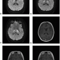
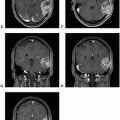
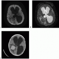
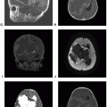
Meningeal melanocytosis is a diffuse tumor that infiltrates leptomeninges but remains within the subarachnoid space. Diffuse enhancement of the leptomeninges on imaging can mimic subarachnoid hemorrhage, pus, or metastatic tumor.
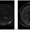
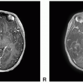
 Figure 7.1. (continued) C. Coronal T1-postcontrast: Avid enhancement of subarachnoid tumor without focal nodularity or mass-like feature. D. Sagittal T1-postcontrast: Avid enhancement of subarachnoid tumor without focal nodularity or mass-like feature. E. Axial T2 FSE: Diffuse subarachnoid tumor is barely visible. F. Axial FLAIR: Slight hyperintensity within the diffuse subarachnoid tumor. G. Diffusion-weighted imaging (DWI): Diffuse subarachnoid tumor is barely visible. H. Apparent diffusion coefficient (ADC): Diffuse subarachnoid tumor is barely visible.
Stay updated, free articles. Join our Telegram channel
Full access? Get Clinical Tree
 Get Clinical Tree app for offline access

|

