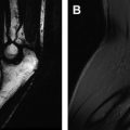
Dr Kimberly Amrami, Professor of Radiology at the Mayo Clinic, Rochester, is spearheading this project as guest editor. Dr Amrami has an excellent knowledge of the subject, including the anatomy, pathology, and technical challenges and details.
A cadre of distinguished radiologists and orthopedic surgeons discusses various important topics regarding MR imaging of the elbow and wrist. Authors include physicians from world-renowned medical centers, including Mayo Clinic, Stanford, Florida Hospital, University of California San Francisco, University of Wisconsin, Brigham and Women’s, University of California Irvine, Favaloro University in Buenos Aires, and Kurume University in Japan.
The content of this issue is timely. New sequences that reduce metal artifact and other techniques, such as isotropic imaging, uTE imaging, T2- and T1-rho mapping, are discussed. Techniques such as MR arthrography, MR angiography, and MR neurography are covered in different articles. Anatomy and pathology in the elbow are succinctly presented. Separate reviews of wrist ligaments, the triangular fibrocartilage, proximal and distal radioulnar joints, carpal fractures, and soft tissue tumors of the upper extremity provide the detail that is needed for today’s imager. In addition, there is a valuable synopsis regarding the relevance of MR imaging to hand surgeons.
Thank you, Dr Amrami and your team of authors, for your hard work to create this exciting and valuable collection of information on these various important topics in the area of MR imaging of the elbow and wrist.
Stay updated, free articles. Join our Telegram channel

Full access? Get Clinical Tree


