KEY FACTS
Terminology
- •
Spectrum of congenital uterine malformations
- ○
20% unicornuate : Single uterine horn ± rudimentary horns
- ○
5% uterus didelphys : 2 separate horns, 2 cervices
- –
Often associated with vaginal septum (75%)
- –
- ○
10% bicornuate : 2 horns with variable degree of fusion
- –
Concave or heart-shaped external fundal contour
- –
May have 1 cervix (bicornis unicollis) or 2 cervices (bicornis bicollis)
- –
- ○
55% septate : Normal, convex external fundal contour
- –
Septum length variable: When complete it extends to external os
- –
- ○
Scanning Tips
- •
Use 3D ultrasound
- ○
Volume acquisition allows reconstruction of true coronal plane to show fundal contour
- –
Septate uterus: Fundus mildly convex to mildly concave
- –
Bicornuate: Concave or heart-shaped external fundal contour
- –
- ○
- •
Look for associated renal anomalies
- ○
Occur in ~ 30% of patients with müllerian duct anomalies
- ○
Unilateral renal agenesis in majority but crossed-fused ectopia, pelvic kidney, horseshoe kidney, renal duplication, and cystic dysplasia all reported
- ○
- •
Look for obstructed component if pelvic pain
- ○
Hematometra: Blood-filled uterus (may involve single horn of duplicated system)
- ○
Hematometrocolpos: Blood-filled uterus and vagina
- ○
- •
Pregnancy failure increased especially with septate uterus
- ○
Use ultrasound guidance for dilatation and evacuation/curettage to ensure appropriate part of cavity is reached
- ○

 adjacent to the pregnancy
  , which is in a unicornuate uterus. Implantation in a rudimentary horn may result in rupture. Color Doppler shows bridging vessels between rudimentary horn & the unicornuate uterus.
 of a uterus didelphys. They are similar in size, excluding a unicornuate uterus with a rudimentary horn. The 2 horns never attach excluding a bicornuate uterus. The pregnancy
  is in the right horn.
 of the didelphys uterus with the pregnancy
  now in the left horn. The cervices are not visible in either image as they are out of the scan plane.
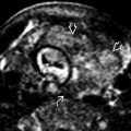
 in the right horn
  of a bicornuate uterus. The empty cavity
  of the left horn is visible at the edge of the picture. Note the cleft
  in the fundal contour, which proves that this is not a septate uterus.
Related posts:


Stay updated, free articles. Join our Telegram channel

Full access? Get Clinical Tree


