KEY FACTS
Terminology
- •
Mechanisms of injury
- ○
Overuse: Strenuous exercise results in diffuse muscular edema
- ○
Stretch: Causes torn muscle fibers + hematoma
- ○
Direct: Usually blunt trauma causes contusion, ± hematoma, ± tear
- ○
Imaging
- •
Delayed-onset muscle soreness
- ○
Overuse leads to diffuse muscular edema
- ○
Muscle may appear normal or diffusely hyperechoic
- ○
- •
Muscle contusion/hematoma
- ○
Contusion: Ill-defined echogenic muscle; dispersed form of hematoma
- ○
Hematoma: Focal hemorrhage often associated with tear
- ○
- •
Muscle or myofascial tear
- ○
Usually partial at/near myofascial junction
- ○
Discontinuity ± retraction of fibers filled with hematoma (hematoma can obscure)
- ○
- •
Muscle hernia
- ○
Focal protrusion of fibers through investing fascia
- ○
Accentuated by contraction or standing
- ○
Clinical Issues
- •
Tears occur at common locations from common mechanisms
- •
Most injuries improve with conservative treatment and do not require follow-up ultrasound
- •
Clinical symptoms overlap with other diagnoses seen with ultrasound (deep vein thrombosis, tendinosis)
Scanning Tips
- •
Use contralateral side for comparison; even if bilateral, abnormalities often appear asymmetric
- •
Carefully assess distal medial gastrocnemius for small tears
- •
Use active contraction/passive joint movement to show retraction of fibers
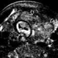
 consistent with delayed-onset muscle soreness. The overlying biceps muscle
  and humeral shaft
  are normal.
 at the myofascial junction
  with the soleus muscle
  . The retraction gap is filled with blood.
Stay updated, free articles. Join our Telegram channel

Full access? Get Clinical Tree


