Musculoskeletal Infections


Musculoskeletal Infections


Infections of the musculoskeletal system can be subdivided into three categories: (a) those involving bones (osteomyelitis), (b) those involving joints (infectious arthritis), and (c) those involving soft tissues (cellulitis). Because of the complexity of the vertebrae and their soft tissue structures, infectious processes of the spine are considered under a separate heading.


INFECTIOUS ARTHRITIS

Three basic mechanisms allow an infectious organism—whether bacterium, virus, mycoplasma, rickettsia, or fungus—to reach the bone: (a) hematogenous spread via the bloodstream from a remote site of infection, such as the skin, tonsils, gallbladder, or urinary tract; (b) spread from a contiguous source of infection, such as from the soft tissues, teeth, or sinuses; and (c) direct implantation, such as through a puncture or missile wound or an operative procedure (Fig. 9.1). Hematogenous spread is common in children, and the usual focus of infection develops in the metaphysis. The metaphyseal location of infection in children is related to an osseous-vascular anatomy that differs in the infant, child, and adult. In the child (ages 1 to 16 years), there is separation of the blood supply to the metaphysis and epiphysis, each having its own source. Moreover, the arteries and capillaries of the metaphysis turn sharply without penetrating the open growth plate; in the region where capillaries become venules, the rate of blood flow is sluggish. Also contributing to the greater incidence of metaphyseal osteomyelitis in children is secondary thrombosis of end arteries with bacteria during transient bacteremia. In the infant (up to 1 year), however, osteomyelitis may sometimes have its focus in the epiphysis because some metaphyseal vessels may penetrate the growth plate and reach the epiphysis. With obliteration of the growth plate in the adult, there is vascular continuity between the shaft and the articular ends of the bone; hence, the focus of osteomyelitis can develop in any part of a bone. Contiguous spread and direct implantation are more common in adults. The sites of bone infection via either of these routes are directly related to the focus of soft tissue infection or the location of the wound.

An infectious agent (Table 9.1) may enter the joint by the same basic routes as in osteomyelitis: by direct invasion of the synovial membrane, either secondary to a penetrating wound or after a joint-replacement procedure, from an infection of the adjacent soft tissues, or indirectly via a blood-borne infection. Infectious arthritis may also occur secondary to a focus of osteomyelitis in the adjacent bone (Fig. 9.2).


Figure 9.1Entry routes of an infectious organism into a bone. Infectious agents may gain entry to a bone through hematogenous spread, a source of infection in the contiguous soft tissues, or through direct implantation secondary to trauma or surgery.


Table 9.1 COMMON ETIOLOGIC AGENTS IN SEPTIC ARTHRITIS


Bacteria


Mycobacteria


Fungi


Spirochetes


Staphylococcus


Mycobacterium tuberculosis


Actinomyces


Treponema pallidum


Diplococcus


Mycobacterium bovis


Coccidioides


Treponema pertenue


Enterococcus


Mycobacterium leprae


Cryptococcus


Borrelia burgdorferi


Serratia


Mycobacterium avium


Histoplasma


Pseudomonas


Mycobacterium ulcerans


Blastomyces


Proteus


Aspergillus


Clostridium


Sporothrix


Bacteroides


Candida


Escherichia


Madurella


Klebsiella


Nocardia


Neisseria


Brucella


PYOGENIC JOINT INFECTIONS


Clinical and Pathologic Features

Pyogenic (septic) arthritis is usually (80% to 90%) monoarticular with predilection for large weight-bearing joints, such as the knee or hip. The clinical signs and symptoms depend on the site and extent of involvement as well as the specific infectious organism. Although most cases of septic arthritis are caused by Staphylococcus aureus, Escherichia coli, and Neisseria gonorrhoeae, other pathogens—including Pseudomonas aeruginosa, Enterobacter cloacae, Klebsiella pneumoniae, Candida albicans, and Serratia marcescens—are being encountered with increasing frequency in joint infections in drug users caused by the contamination of injected drugs or needles. New strains of Staphylococcus aureus may be methicillin (MRSA) or vancomycin resistant. MRSA’s secretion of the necrotizing toxin Panton-Valentine leukocidin results in more invasive infection and higher complication rates. Any large or small joint can be affected by septic arthritis, and hematogenous spread in drug addicts is characterized by unusual locations of the lesion, such as the spine (vertebrae and intervertebral disks), sacroiliac joints, sternoclavicular and acromioclavicular articulations, and pubic symphysis. Furthermore, with the increased usage of biologics and immunosuppressive drugs, clinicians should always keep in mind the possibility that an autoimmune disease has become complicated by an infection. This statement is particularly valid for the patients with rheumatoid arthritis who underwent surgery.


Figure 9.2Entry routes of an infectious organism into a joint. The routes of infection in infectious arthritis are similar to those of osteomyelitis, which itself may be a source of spread.

The classic clinical presentation of septic arthritis is an abrupt, acute onset of joint pain accompanied by swelling and warm sensation. The joint becomes tender to palpation, and there is marked restriction of both active and passive motion. Fever is encountered in about 60% to 80% of cases. Some patients may experience chills. Laboratory findings include elevation of white blood cell (WBC) count, elevation of erythrocyte sedimentation rate, and positive blood cultures. Pathologic findings include progressive and increasing neutrophilic infiltrate of the synovium, associated with vascular dilatation and congestion, and increased number of type A and B synoviocytes. This is followed by cartilage necrosis and subchondral bone erosion. Infiltrates of polymorphonuclear leukocytes on the articular cartilage surface are a common finding (Fig. 9.3). Synovial fluid appears opaque or frankly purulent. The nucleated cell count is very high, up to 50,000 cells per mm3 (see also discussion in Chapter 1). Percutaneous aspiration and US-guided, CT-guided, or fluoroscopy-guided biopsy of a suspected focus of infection may be performed in the radiology suite. It can rapidly confirm a suspected diagnosis of infection and reveal the causative organism. For bacteriologic examination, synovial fluid should be placed into an empty sterile container (not carrying any media) and taken immediately to the laboratory for gram staining
and plating on appropriate media. Chocolate agar (CHOC), Thayer-Martin, or Transgrow media should be used when gonococcal arthritis is considered; Löwenstein-Jensen media, when tuberculous arthritis is suspected; Mannitol salt agar, for staphylococcus infection; and potato dextrose agar or Sabouraud agar for fungi infection.


Figure 9.3Pathology of infectious arthritis. Photomicrograph of a portion of articular cartilage obtained from an acutely inflamed joint shows polymorphonuclear leukocytes on the cartilage surface and underlying erosion of the cartilage (H&E, original magnification ×25). (From Bullough PG. Atlas of Orthopedic Pathology with Clinical and Radiologic Correlation. 2nd ed. New York, NY: Gower Medical Publishing; 1992. Fig. 4.20, p. 4.11.)


Figure 9.4Infectious arthritis. A: A 48-year-old diabetic man presented with pain and soft tissue swelling of the right great toe for the past 3 months. Anteroposterior radiograph shows destruction of the first metatarsophalangeal joint associated with soft tissue swelling and edema typical for septic joint. B: In another patient, a 45-year-old HIV-positive man, who presented with a history of right hip pain for several months, anteroposterior radiograph shows extensive destruction of the right femoral head and right acetabulum. Hip joint aspiration and culture revealed methicillin-resistant Staphylococcus aureus (MARSA) infection.


Imaging Features

In most instances, conventional radiography is sufficient to demonstrate the pertinent features of a bone or joint infection, particularly with the advantage of digital radiography and newest technology of PACS (Picture Archive and Communication System) allowing filmless high-resolution image-display format. Most infectious arthritides demonstrate a very similar radiographic picture, including joint effusion and destruction of cartilage and subchondral bone with consequent joint space narrowing (Fig. 9.4, see also Fig. 3.19). However, certain radiographic features are characteristic of individual infectious processes as demonstrated at various target sites (Table 9.2) and may be helpful in arriving at the correct diagnosis. Generally, a single joint is affected, most commonly a weight-bearing joint like the knee or hip. The early stage of joint infection may be seen simply as joint effusion, soft tissue swelling, and periarticular osteoporosis, but “radiographic” joint space is usually preserved (Fig. 9.5). In the later phase of pyogenic arthritis, articular cartilage is destroyed; characteristically, both subarticular plates are involved and the joint space narrows (Figs. 9.6, 9.7, 9.8, 9.9).

Scintigraphy has a very prominent role in diagnosing musculoskeletal infections, including infectious arthritis. In cases of suspected infection, radionuclide bone scan using


technetium-99m-labeled (99mTc) phosphonates is routinely used, because there is an accumulation of tracer in the infected areas (see Fig. 2.54). A three- or four-phase technique is particularly useful for distinguishing infected joint from infected periarticular soft tissues if radiography is not diagnostic. With cellulitis, diffuse increased uptake is present in the first two phases, but there is no significant increase in uptake in the bone in the third and fourth delayed phases (see Fig. 2.53). Conversely, osteomyelitis causes focally increased uptake in all four phases. The fourphase bone scan can also be useful in diagnosing septic arthritis in situ or with extension into the adjacent bone.


Table 9.2 CLINICAL AND IMAGING HALLMARKS OF INFECTIOUS ARTHRITIS AT VARIOUS TARGET SITES


Type


Site


Crucial Abnormalities


Techniques/Projections


Pyogenic Infections*


Peripheral joints


Periarticular osteoporosis


Joint effusion


Radionuclide bone scan (early)


Standard views specific for site of involvement


Destruction of subchondral bone (on both sides of joint)


Aspiration and arthrography


MRI


Spine


Narrowing of disk space


Loss of definition of vertebral end plate


Anteroposterior and lateral views


Paraspinal mass


CT, MRI


Partial or complete obstruction of intrathecal contrast flow


Myelogram


Destruction of disk


Diskogram and aspiration


Nonpyogenic Infections


Tuberculosis


Large joints


Monoarticular involvement (similar to rheumatoid arthritis)


Radionuclide bone scan


“Kissing” sequestra (knee)


Standard views


Sclerotic changes in subchondral bone


CT


Spine


Gibbous formation


Anteroposterior and lateral views


Lytic lesion in vertebral body


Destruction of disk


Diskogram and aspiration


Paraspinal mass


CT, MRI


Soft tissue abscess (“cold” abscess)


Obstruction of intrathecal contrast flow


Myelogram


Lyme disease


Knee


Narrowing of femoropatellar compartment


Edematous changes in infrapatellar fat pad


Lateral view


CT, MRI


* In IV drug users, unusual sites of infection are encountered, including the vertebra; the sacroiliac, sternoclavicular, and acromioclavicular joints; and the pubic symphysis. The radiologic techniques used to evaluate infections at these sites, as well as the crucial radiographic abnormalities, are the same as those for the more common sites.


Figure 9.5Infectious arthritis. Anteroposterior (A) and lateral (B) radiographs of the left knee of a 4-year-old child demonstrate a significant degree of periarticular osteoporosis and a large joint effusion. Note the small erosions of the distal epiphysis of the femur and the preservation of the joint space. Aspiration revealed hematogenous spread of a staphylococcal urinary tract infection.
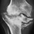


Figure 9.6Infectious arthritis. Anteroposterior (A) and lateral (B) radiographs of the right knee of an 80-year-old man show destruction of articular cartilage of all three joint compartments, erosions of the subchondral bone, posterolateral subluxation, and a large joint effusion.


Figure 9.7Infectious arthritis. Anteroposterior (A) and lateral (B) radiographs of the left knee of a 66-year-old man show destruction of the articular cartilage of all three joint compartments, large erosions of the subchondral bone, posterolateral subluxation, large joint effusion, and soft tissue swelling.


Figure 9.8Infectious arthritis. Oblique (A) and lateral (B) radiographs of the right ankle of a 31-year-old man show destruction of the articular cartilage of the medial malleolus, anterior tibia, and dorsal aspect of the talus, associated with ankle joint effusion.
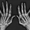


Figure 9.9Infectious arthritis. Radiograph of the toes of a 53-year-old woman shows destruction of the fifth metatarsophalangeal joint associated with a soft tissue swelling and edema.

Once the bone sustains an injury, such as surgery, fracture, or neuropathic osteoarthropathy, that causes increased bone turnover, routine scintigraphy with technetium-labeled phosphonate becomes less specific for infection. However, radionuclide studies using gallium (a ferric analog) and indium are more specific in these instances. There is still no general agreement on the exact mechanism of gallium localization in infected tissues. After intravenous injections of gallium, more than 99% is bound to various plasma proteins, including transferrin, haptoglobin, lactoferrin, albumin, and ferritin. At least five mechanisms of gallium transfer from the plasma into inflammatory exudates and cells have been suggested. These include direct leukocyte uptake, direct bacteria uptake, the protein-bound tissue uptake, increased vascularity, and increased bone turnover. Because gallium binds to the iron-binding molecule transferrin, the mechanism of gallium uptake in infectious processes is best explained by hyperemia and elevated permeability that increase delivery of
the protein-bound tracer transferrin into the area of inflammation. Cells associated with the inflammatory response, particularly polymorphonuclear white cells in which lactoferrin is carried within intracytoplasmic granules, deposit iron-binding proteins extracellularly at the site of inflammation, serving to combat the infection by sequestering needed iron from bacteria. Lactoferrin, which has a high binding affinity for iron, takes the gallium away from the transferrin.

The other tracer used in infections is indium. Because indium-labeled white blood cells are usually not incorporated into areas of increased bone turnover, scintigraphy with indium-111 (111In) oxine-labeled leukocytes is used as a sensitive and specific test in the general diagnosis of infection of the musculoskeletal system and in specific instances when infection complicates previous fracture or surgery. Like other imaging procedures in nuclear medicine, this test monitors the internal distribution of a tracer agent to provide diagnostic information. The inherent ability of white blood cells to accumulate at sites of inflammation makes their use in this test particularly effective in the diagnosis of infections (Fig. 9.10, see also Fig. 2.62). Merkel reported the sensitivity of indium scintigraphy in detecting infections to be 83%,
with a specificity of 94% and an accuracy of 88%. It must be stressed, however, that because the 111In-labeled leukocytes also accumulate in active bone marrow, the sensitivity for the detection of chronic osteomyelitis is reduced. To improve the diagnostic ability of this technique, a combined 99mTc-sulfur colloid bone marrow/111In-labeled leukocyte study is advocated. A particularly difficult problem is the patient with diabetic foot neuropathy in whom superimposed infection is suspected. In this circumstance, radiography and even MRI are not very specific. Although soft tissue infection can be detected by the latter technique, early changes of osteomyelitis and septic arthritis may be missed. Often, no single imaging method can provide the correct diagnosis, and a combination of several imaging techniques should be used. The traditional sequential use of 67Ga citrate in conjunction with the 99mTc-methylene diphosphonate (MDP) bone scan as an aid to diagnose osteomyelitis and infectious arthritis in the diabetic foot has been supplanted in recent years by the use of 111In-labeled leukocytes. The drawback of this technique is that there remain difficulties in differentiating infection in the bone (osteomyelitis) from that in the adjacent tissue (cellulitis). A more recent attempt to improve this situation is the use of a combined 99mTc-bone scan/111In-labeled leukocyte study to determine whether the leukocyte collection is in the bone or in the soft tissue. A new challenger to 111In leukocyte scanning is the 99mTc-hexamethylpropylene amino oxine (HMPAO)-labeled leukocyte scan. At the time of this writing, other methods are being tested, namely, isotope-labeled (99mTc, 111In, or 123I) monoclonal antigranulocyte antibodies, isotope-labeled polyclonal IgG, isotope-labeled monocytes, isotope-labeled chemotactic polypeptide analogs, and isotope-labeled specific antibodies against bacteria.


Figure 9.10Scintigraphy and MRI of infectious arthritis. A: Anteroposterior radiograph of the left shoulder of a 20-year-old woman shows moderate periarticular osteoporosis, osteolytic ill-defined lesions in the glenoid and proximal humerus, and periosteal reaction at the lateral aspect of the humeral shaft. B: 111In oxine-labeled white blood cell scintigraphy shows increased uptake of the radiopharmaceutical agent in the right shoulder (arrow). Coronal (C) and axial (D) T1-weighted fat-suppressed MR images obtained after administration of gadolinium show significant enhancement of the bones and surrounding soft tissues. The aspiration/biopsy followed by bacteriologic examination revealed Bacteroides fragilis.

Over the past decade, FDG-PET/CT has emerged as a rapidly evolving diagnostic tool for evaluation of infections. In particular, recent studies by Diaz and collaborators using a combination of [124I] FIAU-positron emission tomography and CT demonstrated positive signals at the site of infection at two hours after administration of 2mCi (74 MBq) of radiopharmaceutical agent. FIAU [1-(2′-deoxy-2″fluoro-beta-D-arabinofuranosyl)-5-iodouracil], a nucleoside analog that freely enters and exits cells, is a substrate for the native thymidine kinase (TK) from a wide variety of bacteria. Once phosphorylated by TK, [124 I] FIAU becomes trapped within bacteria and can be detected with PET/CT.


Figure 9.11CT of osteomyelitis and infectious arthritis. Axial (A), coronal reformatted (B), and sagittal reformatted (C) CT images of the left foot of a 72-year-old diabetic man demonstrate an active osteomyelitis of calcaneus and infection of the subtalar joint. Note several sclerotic osseous fragments representing sequestra (arrows).

Arthrography has rather limited application in the diagnosis of joint infections. This radiologic technique, which is often performed after aspiration of the joint to obtain a fluid specimen for bacteriologic examination, helps determine the extent of joint destruction and demonstrate the presence of synovitis (see Fig. 2.30). CT plays a determining role in demonstrating the extent of infection in bones and joints (Figs. 9.11 and 9.12). Ultrasound (US) can occasionally be used in diagnosing soft tissue and joint infections, as well as osteomyelitis. This modality has the advantage of being easily accessible and available at relatively reasonable cost. In addition, this technique does not expose the patient to ionizing radiation. Real-time capability of US is unique in providing a means to evaluate structures under dynamic conditions. Furthermore, US plays an important role in the guidance of percutaneous biopsy and aspiration of infectious lesions, as well as the therapeutic drainage of abscesses.

At the present time, MRI established its place in the evaluation of musculoskeletal infections. As several studies have indicated, osteomyelitis, soft tissue abscesses, joint and tendon sheath effusions, and various forms of cellulitis are well depicted by this modality. MRI is as sensitive as 99mTc-MDP in demonstrating osteomyelitis and infectious arthritis and more sensitive and more specific than other scintigraphic
techniques in demonstrating soft tissue infections, primarily because of its superior spatial resolution. The proper evaluation of musculoskeletal infections with MRI requires both T1- and T2-weighted images in at least two imaging planes. In anatomically complex areas such as the pelvis, spine, foot, and hand, three planes may be necessary. MRI manifestations of pyogenic arthritis include joint effusion with surrounding soft tissue edema and bone marrow edema (Figs. 9.13 and 9.14). In more advanced stages, cartilage and bone destruction may be seen, due to associated osteomyelitis (Figs. 9.15, 9.16, 9.17, see also Fig. 9.4). “Lamellated” joint effusion demonstrated with MRI has been described as a reliable sign of septic arthritis.
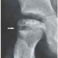


Figure 9.12CT of infectious arthritis. A 31-year-old IV drug abuser presented with anterior chest pain and fever. A: Lateral radiograph of the chest shows a large soft tissue swelling anteriorly to the upper sternum (arrow). B: Sagittal CT image of the chest demonstrates erosive changes of the manubriosternal joint (arrow). C: Coronal reformatted CT image shows more clearly the joint erosions (arrows).
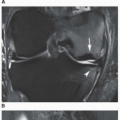


Figure 9.13MRI of septic arthritis. Coronal T2-weighted MRI of the right hip in a 12-year-old boy demonstrates a joint effusion with capsular distension (arrow). There is edema of the surrounding muscles. There are no signs of osteomyelitis. The arthrocentesis and bacteriologic studies of the obtained fluid confirmed the presence of joint infection.


Figure 9.14MRI of septic arthritis. A: Radiograph of the index finger of a 22-year-old man shows narrowing of the proximal interphalangeal joint, periarticular osteoporosis, and soft tissue swelling. B: Axial T1-weighted MR image shows low-intensity signal of the bone marrow and surrounding soft tissues of the index finger (compare with normal middle and ring fingers). Axial (C) and coronal (D) T1-weighted fat-suppressed MR images obtained after intravenous administration of gadolinium show significant enhancement consistent with infectious synovitis, cellulitis, and osteomyelitis.


Figure 9.15MRI of septic arthritis. A: Dorsovolar radiograph of the right wrist of a 43-year-old man shows destruction of the radiocarpal joint and erosive changes of the distal radius, distal ulna, lunate, and scaphoid bones. Note also involvement of the carpometacarpal articulation. There is periosteal reaction of the distal radius and ulna and soft tissue swelling. B: Coronal 3D GRE fat-suppressed and coronal proton density-weighted fat-suppressed MR images demonstrate an erosion of the distal ulna (arrow) with a radiocarpal joint effusion extending to the distal radioulnar joint through a complete tear of the triangular fibrocartilage complex. Note the intermediate-to-low signal intensity of most of the effusion and mild surrounding soft tissue edema (arrowheads) consistent with synovitis due to septic arthritis.
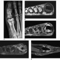


Figure 9.16MRI of septic arthritis. A: Dorsoplantar radiograph of the right forefoot of a 52-year-old man shows destruction of the first metatarsophalangeal joint, erosions of the head of the first metatarsal bone, and a large soft tissue swelling. Short-axis (B) and sagittal (C) T1-weighted MR images show low-intensity signal of partially destroyed head of the first metatarsal bone and surrounding soft tissues (arrows). Short-axis (D) and sagittal (E) T1-weighted fat-suppressed MR images obtained after administration of gadolinium show enhancement within the joint and bone marrow (arrows) indicative of joint infection and osteomyelitis.


NONPYOGENIC JOINT INFECTIONS


Tuberculous Arthritis

Musculoskeletal tuberculosis accounts for 1% to 3% of tuberculous infections, and most commonly results from

hematogenous or lymphatic dissemination of mycobacterium. Tuberculous arthritis represents 1% of all forms of extrapulmonary tuberculosis, although the number of cases has recently been on the rise. The acid-fast tubercle bacilli Mycobacterium tuberculosis and Mycobacterium bovis are the causative organisms. The infection may be found in all groups, but more commonly in children and young adults. Predisposing factors such as trauma, alcoholism, drug abuse, intra-articular injection of steroids, or prolonged systemic illness are found in most patients with tuberculous arthritis. The joint infection usually is caused by either direct invasion from an adjacent focus of osteomyelitis or hematogenous dissemination of the tubercle bacillus. Large weight-bearing joints such as the hip or knee are most often affected, and monoarticular involvement is the rule. Involvement of the small joints of the hands and feet may be observed in children and immunocompromised patients.


Figure 9.17CT and MRI of septic arthritis. Anteroposterior (A) and lateral (B) radiographs of the right ankle of a 65-year-old man show narrowing of the ankle joint, erosions of the distal tibia and talus, ankle joint effusion, and soft tissue swelling. Coronal (C) and sagittal reformatted (D) CT images show destructive joint changes more effectively. In addition, there are several small bone fragments within the joint space representing sequestra. Observe also periosteal reaction.


Figure 9.17 ▪ (Continued) Sagittal T1-weighted (E), inversion recovery (IR) (F), and T1-weighted fat-suppressed (G) postcontrast MR images show typical changes of septic joint and osteomyelitis.


Clinical and Pathologic Features

Most common findings include joint pain and swelling, decreased active and passive movement in the joint, lowgrade fever, and excessive sweating, especially at night.

Histopathologic examinations, culture identification, and polymerase chain reaction (PCR) are among the most accurate methods for diagnosis of tuberculosis. Gross pathologic examination of the areas affected by tuberculosis shows thickened edematous tissue, commonly studded with small grayish nodules, exhibiting white opaque center (granulomas). They may become confluent and produce larger areas of white necrotic material representing caseation (cheesy) necrosis. In the joint, separation of the articular cartilage dissected from underlying subchondral bone by granulomatous tissue is a characteristic feature. On microscopic examination, the typical tubercle consists of a central necrotic area surrounded by pale epithelioid histiocytes. Among these cells noted are scattered giant cells with nuclei typically arranged at the periphery of the cell, known as Langhans giant cells (Fig. 9.20). At the periphery of the tubercle, there is a rim of mixed chronic inflammatory cells present.

Only gold members can continue reading. Log In or Register to continue

Stay updated, free articles. Join our Telegram channel

Oct 4, 2018 | Posted by in GENERAL RADIOLOGY | Comments Off on Musculoskeletal Infections

Full access? Get Clinical Tree

Get Clinical Tree app for offline access