Abstract
Objective
Near-field (NF) clutter filters are critical for unveiling true myocardial structure and dynamics. Randomized singular value decomposition (rSVD) stands out for its proven computational efficiency and robustness. This study investigates the effect of rSVD-based NF clutter filtering on myocardial motion estimation.
Methods
In silico , material points and their displacements in a homogeneous medium under uniaxial compressions (0.5% – 9% axial strains at 0.5% increments) were simulated in finite-element models. They were exported to the k-Wave toolbox for simulations of pre- and post-deformed ultrasound images with/ without a realistic phase aberrating layer in a high-contrast diverging wave compounding scheme. In vivo , echocardiograms of 20 normal human hearts were acquired using a coded diverging wave compounding imaging method at 3200 frames/second in the transthoracic apical four-chamber view. Morphological component analysis (MCA), which is also a sparse representation method but computationally intensive, was used for comparison with rSVD. Both rSVD- and MCA-based filters were applied to beamformed ultrasound radio-frequency (RF) data before cross-correlation-based speckle tracking. Contrast-to-noise ratios (CNRs) and root-mean-square deviations (RMSDs) were computed from regions of interest to evaluate NF clutter filtering performance of rSVD and MCA.
Results
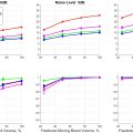
In silico , 2-D displacements estimated from rSVD-based clutter-reduced image data showed strong agreement with ground truth (R 2 of 0.95). In vivo , CNR improvements ranged from 1.02 dB to 17.68 dB, consistently enhancing image quality across all subjects. An improvement of ∼4.9 dB in the apical segments was observed in 80% of cases. Mean RMSDs were below 5.0% for all rSVD-based NF clutter-reduced data. While both rSVD and MCA effectively filtered NF clutter, rSVD was significantly more practical.
Conclusion
Our findings confirm the reliability, accuracy, and efficiency of rSVD-based clutter filtering in speckle tracking echocardiography. This underscores the feasibility of matrix decomposition-based methods, exemplified by rSVD, in NF clutter filtering for myocardial motion estimation.
Introduction
Speckle tracking echocardiography is a well-established technique for assessing myocardial kinematics [ ]. Abnormal wall motion indicates myocardial impairment from myocardial ischemia or disrupted conduction [ , ]. However, near-field (NF) clutter, originating from multi-path reverberations or off-axis scatterers, degrades echocardiographic images, appearing as a high-level stationary noise in the upper field of view (FOV) and diminishing with depth [ ]. Near-field clutter complicates visual scoring and causes myocardial motion estimation inaccuracies [ ], leading to diagnostic uncertainties. Filtering NF clutter is thus imperative to unveil the actual myocardial motion, especially in the apical region, where myocardial ischemia risk is high [ , ].
An early work introduced a temporal high-pass filter to reduce NF clutter [ ]. This filter was applied to beamformed radio-frequency (RF) signals at specific spatial locations over consecutive frames, using a 15 Hz cutoff to prevent tissue information loss. Zwirn and Akselrod [ ] later proposed a clutter detection algorithm, hypothesizing that NF clutter exhibited subtle pixel-wise spatial grey-level changes within a cardiac cycle. They categorized histograms of mean intensity values and root mean square errors into Gaussian-modeled sections: nearly stationary, low-velocity, and high-velocity pixels [ ], setting thresholds at Gaussian functions intersections. The performance of their method depended heavily on parameter selections and risked misidentifying small bright regions as clutter.
Matrix decomposition-based methods were thereafter applied to echocardiography NF clutter reduction. Lediju et al . [ ] used principal component analysis (PCA) to show NF clutter was nonstationary. Mauldin et al . [ ] developed a singular value filter (SVF), utilizing weighted orthogonal basis functions from eigenvectors and singular value spectra of windowed beamformed RF or in-phase quadrature (IQ) signals. Compared with conventional PCA and high-pass filters, SVF demonstrated superior clutter rejection and motion estimation in in vivo murine echocardiographic data, with higher contrast-to-noise ratios (CNRs) and displacement signal-to-noise ratios (SNRs). However, SVF may attenuate myocardial signals in clutter-free areas. Meanwhile, Turek et al . [ ] proposed morphological component analysis (MCA) to sparsely represent tissue and clutter signals; this preserved tissue signals and achieved CNR improvement comparable to SVF in human heart data. However, the effect of MCA on speckle tracking was not investigated [ ].
Despite their excellent performance, SVD-based and MCA-based methods require substantial computing resources. To reduce computational complexity, a research group proposed closed-loop low-rank echocardiographic artifact removal (CLEAR) [ ], which used a “random sampling with replacement” method to detect clutter and removed it by an SVF-based method. This global processing approach was validated using in vivo mouse heart datasets with synthetic clutter, showing that CLEAR reduced tracking errors by approximately 50%, performing comparably to MCA but better than SVF.
Matrix decomposition-based methods assume NF clutter is nearly stationary, but myocardium can be quasi-stationary during diastasis. To address this issue, Sjoerdsma et al. [ ] developed a frame-by-frame method that does not rely on any particular motion assumptions. They employed complex-valued oriented fast wavelets to suppress NF clutter present in the highest and lowest subbands. This method is agnostic to temporal factors and preserves ultrasound speckles, thus rendering it suitable for speckle tracking tasks.
The aforementioned filters rely on linear representations of data. Learning-based methods can learn global spatiotemporal features and model non-linear systems. Tabassian et al. [ ] trained a 3-D U-Net to remove synthetically superimposed artifacts in synthetic 2-D echocardiograms, outperforming the SVD-based clutter filter proposed in [ ]. However, its generalizability to in vivo data was not demonstrated.
Currently, SVD-based filters are widely used for in vivo ultrasound imaging but are constrained by computational complexity and their dependency on spatial and temporal window selection [ ]. Randomized SVD (rSVD), developed by Halko et al. [ ] and optimized by Martinsson PG [ ], overcomes these limitations and exhibits high accuracy, robustness, and computation speed. Song et al. [ ] and Lok et al. [ ] validated the accuracy and real-time capability of rSVD primarily in micro-vessel ultrasound imaging applications, where tissue motion is often minimal, and the algorithmic robustness is thus less critical. In contrast, we applied rSVD in echocardiography [ ], where tissue motion is highly dynamic, to demonstrate the robustness of rSVD under challenging conditions through the validation of our hypothesis that SVD performance is closely linked to the tissue velocity gradient. Although Sjoerdsma et al. [ ] have investigated speckle preservation using a spatial frequency domain-based filter, the reliability of filtered signals from linear decomposition methods remains unexplored. By efficiently capturing matrix characteristics without requiring spatial and/or temporal window selection, rSVD serves as an example of a linear matrix decomposition method well-suited for this purpose. Hence, we propose rSVD for NF clutter reduction in echocardiography in vivo and evaluate the reliability of filtered signals for subsequent physiological motion estimation.
Despite the significance of in vivo demonstrations, the unavailability of ground truth poses a challenge, making in silico studies indispensable for post-filtered signal analysis. However, simulations in previous studies either superimposed unrealistic artifacts [ – , ] or only considered axial translation of the medium [ – ], neglecting both multi-path reverberations and medium deformations. Additionally, whether the random scheme in rSVD impacts the accuracy of filtered ultrasound signals for 2-D speckle tracking has not been reported.
In our in silico study, a defined scatterer field first underwent 2-D (axial and lateral) deformation in a finite-element (FE) model. Simulated NF clutter arose from ultrasound wave propagation through anatomically realistic tissue layers above the deforming medium. Whether the preserved speckles could reveal real motion patterns was validated against the ground truth motion from the FE model. The efficacy of rSVD for NF clutter reduction and myocardial motion estimation was assessed in an in vivo study on human hearts, and further compared to the filtering performance of MCA, a well-established but computationally intensive sparse representation method, as mentioned earlier.
Method
Principle of randomized singular value decomposition (rSVD)
The randomized scheme in rSVD [ , ] efficiently identifies the most significant singular vectors. However, when singular values decay slowly, distinguishing between primary and secondary components becomes difficult. Power iteration helps mitigate this issue by amplifying the dominant singular values, thus easily highlighting the dominant singular vectors for effective component separation [ , ]. This is especially important for cross-correlation-based motion tracking algorithms on filtered data. Key steps in implementing the power iteration scheme are described, with details in [ ].
After reshaping the 3-D ultrasound data matrix (regardless of the signal format: channel, aperture or beamformed; RF or IQ data) matrix into a 2-D matrix A ( Figures 1 a, b), matrix A is processed through q power iterations to create matrix H
where * denotes complex conjugate transpose, and q is an integer. The singular values decay more rapidly as σ j ( H ) = σ j ( A ) 2 q + 1 , where σ j represents the j -th singular value. As additional iterations significantly increase computational costs without providing proportional gains in accuracy, q = 1 or q = 2 usually suffices.

In this study, the first power iteration ( i.e. , q = 1 ) adequately balanced computational efficiency and accuracy across our data, as demonstrated in Figure 1 c. To delineate the subspace boundary between NF clutter and myocardial signals, singular value thresholding was determined by the spatial similarity matrix [ ]. An initial threshold k 0 , was empirically set at the first 10% of the rank of the 2-D matrix A , with the optimal threshold k illustrated in Figure 1 d. The robust rSVD filter requires only one single threshold for all frames in a cardiac cycle.
Finite-element simulation
A 3-D homogeneous and isotropic phantom (20 × 20 × 20 mm 3 ) was simulated using Abaqus/CAE (6.12-1, Dassault Systems, Veliz-Villacoublay, France) [ ]. The phantom, linearly elastic with a Young’s modulus of 50 kPa and incompressible (Poisson’s ratio = 0.499), underwent stepwise uniform compression, with axial strains increasing from 0 % to 9 % in 0.5 % increments. Axial downward motion was applied at the top surface, while the bottom surface was fixed. Nineteen deformation states were individually incorporated into the ultrasound simulation scheme.
2-D ultrasound phased-array image simulation
The k-Wave MATLAB toolbox [ ] was used to simulate 2-D ultrasound images of the pre- and post-deformed FE phantom. A phased array probe, modeled after the P4-2 probe (Verasonics, Kirkland, WA, USA) used in in vivo experiments, was positioned at the top of the FOV. Received signals were beamformed and coherently compounded [ ]. Table 1 lists the simulation parameters.
| Grid size | 15 μm |
|---|---|
| Simulation region size (ordinate × abscissa) | 2668 × 3334 grids (40 × 50 mm 2 ) |
| PML size | 20-grid |
| Time step size | 2.5 ns |
| Acquisition time (each transmission) | 72.13 μs |
| CFL number | 0.3 |
| Sound velocity | 1540 m/s |
| Probe type | 64-element phased array |
| Pitch | 0.32 mm |
| Element width | 0.3 mm |
| Center frequency | 2.5 MHz |
| Type of emission | diverging wave |
| Angular aperture | 90° |
| Subaperture number | 2 |
| Emission elements | 32 |
| Reception elements | 64 |
| Simulation platform | Linux system with 40 CPU cores (Intel(R) Xeon(R) Gold 6138 CPU at 2.00GHz) and 124.5 GiB memory. |
Material points and their in-plane displacements in the central depth-width slice of the FE phantom were exported, with out-of-plane motion excluded. The 2-D slice (20 × 20 mm 2 ) was centered and placed in the imaging depth between 15 mm and 35 mm of the k-Wave computational domain. The scattering field comprised both the background medium and the FE phantom. Figure 2 shows spatial distributions of the speed of sound and density, comparable to those of human soft tissues [ , ]. A 2-D heterogeneous tissue modeled by Mast et al . [ ] was adopted to mimic in vivo NF clutter, comprising layers of fat, skeletal muscle, connective tissue, and skin. Reference values of acoustic properties were summarized in Table 2 .

| Tissue | c 0 (m/s) | ρ (kg/m 3 ) | B / A | α 0 (dB/MHz/cm) | y |
|---|---|---|---|---|---|
| Background | 1540 | 1000 | 8.0 | 0.30 | 1.5 |
| Fat [ ] | 1479 | 937 | 9.6 | 0.40 | |
| Skeletal muscle [ , ] | 1547 | 1050 | 6.6 | 0.74 | |
| Cardiac muscle [ , ] | 1545 | 1060 | 7.1 | 0.52 | |
| Connective tissue [ ] | 1613 | 1120 | 8.0 | 0.68 | |
| Skin [ ] | 1615 | 1090 | 7.9 | 0.35 |
In vivo echocardiography
Twenty healthy subjects (10 males and 10 females; 72 ± 5 y.o.) with a BMI of 17.1-27.4 were recruited in this study, approved by the Institutional Review Board of the University of Hong Kong (UW19-043). Informed consent was given and signed.
Echocardiography of the apical four-chamber view was performed using a Vantage 256 system with a P4-2 probe ( f c = 2.5 MHz) and our previously developed cascaded synthetic aperture (CaSA) imaging method [ ] at 400 compounded frames/second. The array was divided into eight subapertures on transmit, each emitting eight encoded cascaded diverging waves. In reception, the entire array aperture was used. High SNR images were achieved by decoding backscattered signals, and coherent compounding was performed after delay-and-sum beamforming. The electrocardiogram (ECG) was synchronously recorded using a BIOPAC system (MP36, Goleta, CA, USA).
2-D RF-based speckle tracking
A previously developed 2-D cross-correlation-based speckle tracking method [ ] was used to estimate the 2-D in-plane motion between successive beamformed RF frames. Table 3 lists speckle tracking parameters. Strains were then computed using a least-squares strain estimator (LSQSE) [ ], with kernel sizes of 6 mm and 10 mm for the axial and lateral displacement maps, respectively.
| Scenario | ||
|---|---|---|
| Parameter | In silico | In vivo |
| Axial kernel size (pre-recorrelation) | 4λ (=2.46 mm) | 10λ (=6.16 mm) |
| Lateral kernel size (pre-recorrelation) | 12 d p (=3.84 mm) | 1 d p (=0.32 mm) |
| Axial kernel size (post-recorrelation) | 4λ (=2.46 mm) | 10λ (=6.16 mm) |
| Lateral kernel size (post-recorrelation) | 12 d p (=3.84 mm) | 7 d p (=2.24 mm) |
| Axial kernel overlap | 80% | 80% |
| Lateral kernel shift | 1 d p | 1 d p |
| Upsample factor (spline interpolation in the lateral direction) | 1 | 8 |
Performance evaluation
The NF clutter reduction performance was evaluated by two metrics. The first metric was the CNR, defined as
where μ T and μ B are the mean intensities of the target and background, respectively, and σ T and σ B are the intensity standard deviations (SDs) of the target and background, respectively. In the in vivo study, differential CNRs (ΔCNRs) ( i.e., CNR clutter reduction -CNR original ) [ ] were calculated throughout the cardiac cycle from specific myocardial segments.
The second metric assessed was motion estimation accuracy. In silico , linear regression and Bland-Altman analysis [ ] were used to evaluate the agreement between motion estimates and ground truth values with a determination coefficient of 0.95. In vivo , the root-mean-square deviation (RMSD) was computed as
Related posts:
 Advances in the Application of Artificial Intelligence in the Ultrasound Diagnosis of Vulnerable Carotid Atherosclerotic Plaque
 Changes in Muscle Quality Following Short-Term Resistance Training in Older Adults: A Comparison of Echo Intensity and Texture Analysis
 Guided-VCTE: An Enhanced FibroScan Examination With Improved Guidance and Applicability
 Regional Image Quality Scoring for 2-D Echocardiography Using Deep Learning
 Ultrafast Coherence-Based Power Doppler Estimation Using Nonlinear Compounding With Complementary Subset Transmit
 Characterization of LIPUS Parameters Suitable for Hip Bone Fracture
Stay updated, free articles. Join our Telegram channel

Full access? Get Clinical Tree


