Abstract
Hemophagocytic lymphohistiocytosis (HLH) is a severe condition characterized by the secretion of large amounts of inflammatory cytokines. Lymphoma is a major cause of secondary HLH. This report describes the case of a child initially diagnosed with HLH who experienced recurrent episodes after treatment. Ten months after the initial diagnosis, a mass was discovered in the right upper limb, and pathology findings confirmed NK/T-cell lymphoma. Based on the patient’s medical history, this lymphoma was considered the underlying cause of HLH. Patients diagnosed with lymphoma complicated by HLH have a worse prognosis and shorter survival compared with those without HLH. Early diagnosis and timely symptomatic treatment can significantly improve patient prognosis.
Introduction
Hemophagocytic lymphohistiocytosis (HLH) is a clinical syndrome characterized by a pathological inflammatory response resulting from inherited or acquired abnormalities in immune function [ ]. HLH occurring during the diagnosis and treatment of lymphoma is collectively referred to as lymphoma-associated HLH (LA-HLH). In cases caused by lymphoma itself, HLH may occur before, simultaneously with, or at the relapse or progression of lymphoma [ ]. High levels of inflammatory factors, such as interleukin-6 (IL-6) and interferon-γ (IFN-γ), secreted by lymphoma cells, may contribute to the development of HLH [ ]. This type of HLH is most commonly seen in NK/T-cell lymphoma. In this case, a child was initially admitted to the hospital with a diagnosis of HLH. After treatment, the condition recurred, and the child was readmitted with a mass in the right upper extremity. Pathological examination confirmed NK/T-cell lymphoma. Retrospectively, the entire clinical course was considered consistent with LA-HLH, and the details are reported below.
Case presentation
A boy aged 2 years and 4 months was initially admitted to the hospital due to fever lasting 15 days. The fever ranged from 38°C-39°C, peaking at 40.3°C, with no clear pattern. The child had chills, an occasional cough, and a runny nose. Laboratory tests showed a white blood cell (WBC) count of 5.03 × 10 9 /L (reference range: 5-12 × 10 9 /L), neutrophil percentage of 32.6% (reference range: 40%-75%), red blood cell (RBC) count of 3.84 × 10 12 /L (reference range: 3.5-5.5 × 10 12 /L), hemoglobin (Hb) of 98 g/L (reference range: 110-160 g/L), and platelet count (PLT) of 81 × 10 9 /L (reference range: 100-300 × 10 9 /L). The child received cefoperazone combined with azithromycin for anti-infective therapy.
One day later, WBC decreased to 3.59 × 10 9 /L, neutrophil percentage was 32.6%, RBC was 3.64 × 10 12 /L, Hb 92 g/L, and PLT 46 × 10 9 /L. Due to decreased WBC, Hb, and PLT counts, a hematological malignancy could not be excluded. Bone marrow cytology was performed for further diagnosis, revealing visible phagocytosis. Bone marrow examination confirmed hemophagocytosis, and ferritin was elevated to 1045.5 ng/mL (reference range: 20-250 ng/ml). The child was clinically diagnosed with hemophagocytic syndrome and treated with intrathecal chemotherapy (dexamethasone and methotrexate) via lumbar puncture. After 17 days, laboratory results showed WBC 6.21 × 10 9 /L, Hb 98 g/L, PLT 780 × 10 9 /L (above the reference range), and ferritin 289.2 ng/mL. Repeated bone marrow cytology no longer showed phagocytic cells and appeared normal. The child was subsequently discharged.
Over the following months, he was repeatedly hospitalized for fever and continued the previous chemotherapy regimen, which alleviated symptoms.
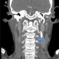
Ten months after disease onset, the child presented again with fever and a palpable subcutaneous mass (approximately 30 mm × 20 mm) in the right upper arm. The mass was slightly hard, without redness, swelling, or tenderness. Red papules, approximately 2-8 mm in diameter, appeared scattered on the skin, mainly on the limbs. Soft-tissue ultrasound showed a hypoechoic mass in the subcutaneous muscle tissue of the right upper arm, and a hematoma could not be excluded. Laboratory results on admission were: WBC 4.2 × 10 9 /L (slightly below reference range), neutrophil percentage 24.6% (below reference range), Hb 98 g/L (below reference range), PLT 216 × 10 9 /L (within reference range), lymphocyte percentage 51.7% (elevated, reference range: 20%-40%). X-ray (frontal and lateral views) of the right humerus revealed a small periosteal reaction in the lower humerus, suggestive of possible osteochondritis dissecans ( Fig. 1 ). Anti-infective treatment was administered but was ineffective. CT scan of the right upper arm also showed a small periosteal reaction in the lower humerus ( Fig. 1 ). Soft tissue biopsy indicated malignant proliferation of NK/T cells in the right elbow region. MRI with enhancement of the right upper arm revealed bone destruction in the middle and lower humerus accompanied by a soft tissue mass, consistent with lymphoma ( Fig. 2 ).

Related posts:
 A rare and life-threatening case of spontaneous hemopneumothorax presenting with severe hemodynamic instability
 A very rare case of ileocolic and appendiceal intussusception with acute appendicitis
 Hepatic and extra-hepatic hydatid cysts: A case series of radiological and clinical insights
 Mid-term follow-up of a fractured flow diverter in the internal carotid artery
 An atypical multiple autoimmune syndrome: A case report including myocarditis
 Persistence of the hypoglossal artery: A rare case report
Stay updated, free articles. Join our Telegram channel

Full access? Get Clinical Tree


