Nuclear Cardiology
QUESTIONS
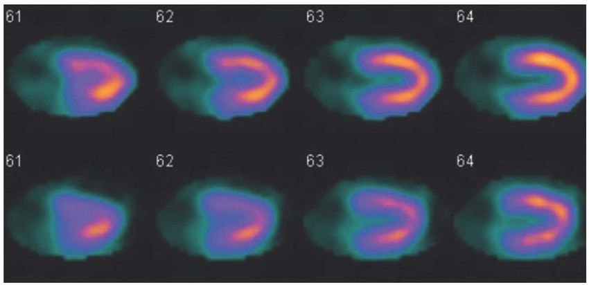
1 The following image is from a dual-isotope myocardial perfusion examination. What is the major difference between performing the myocardial perfusion stress imaging with thallium-201 (Tl-201) and technetium-99m (Tc-99m) myocardial perfusion agents?
 |
A. Higher dose of thallium-201 is used for imaging.
B. Stress acquisition must begin within 10 minutes of Tl-201 injection.
C. Exercise stress test cannot be performed with Tl-201.
D. Pharmacologic stress test cannot be performed with Tl-201.
E. Imaging is usually begun 30 to 45 minutes after Tl-201 administration.
F. Tl-201 has less soft tissue attenuation.
View Answer
1 Answer B. Thallium-201 (Tl-201) is a potassium analog, which is transported across the myocardial membrane via the Na+-K+ ATPase pump into the myocardial cytosol. It has a very high first-pass extraction rate of approximately 85% compared to about 60% for the Tc-99m-labeled myocardial perfusion imaging agents (sestamibi or tetrofosmin). Unlike Tc-99m-labeled myocardial perfusion imaging agents, Tl-201 quickly undergoes redistribution, which is a dynamic exchange of Tl-201 between the myocardial cytosol and the vascular blood pool. Because of redistribution, poststress images with Tl-201 must be performed quickly after its injection (<10 minutes). Redistribution is also the basis for the use of Tl-201 in the evaluation of myocardial viability with delayed 24-hour imaging. Either exercise or pharmacologic stress protocol can be used with Tl-201. Because of its higher absorbed dose, in part due to its longer half-life of 3 days, the injected dose of the Tl-201 is lower than the Tc-99m-labeled myocardial perfusion agents. Biliary excretion of Tc-99m-labeled myocardial perfusion agents results in intense activity within the liver. In order to allow clearance from the liver and since these agents do not undergo redistribution, imaging is usually begun 30 to 45 minutes after their administration. Since Tc-99m photon has higher energy (140 keV) than mercury daughter x-rays from Tl-201 (69 to 81 keV), less soft tissue attenuation would be seen with Tc-99m-labeled agents compared to Tl-201.
Reference: Mettler FA, Guiberteau MJ. Essentials of nuclear medicine imaging, 6th ed. Philadelphia, PA: Saunders, 2012:135-140.
2 What is the major difference between the PET myocardial perfusion images acquired with N-13 ammonia when compared to rubidium-82?
A. Better resolution
B. Larger full width at half-maximum
C. No difference
D. Higher energy photon
E. Less signal to noise
View Answer
2 Answer A. N-13 ammonia positron has less energy than the positron emitted by the decay of rubidium-82 (Rb-82). As such, it travels shorter distance before undergoing annihilation. This decreased range is responsible for the better image resolution (smaller full width at half-maximum) of N-13 ammonia myocardial perfusion images compared to that acquired with Rb-82. The photons generated by annihilation are 511 keV for both N-13 ammonia and Rb-82. N-13 ammonia, Rb-82, and F-18 flurpiridaz are the PET radiotracers that can be used to evaluate myocardial perfusion; F-18 FDG is used for the evaluation of myocardial viability. The use of N-13 ammonia (half-life 10 minutes) requires an on-site cyclotron. Rubidium-82 (half-life 76 seconds) is produced by a generator and does not require an on-site cyclotron. F-18 flurpiridaz is a new PET myocardial perfusion radiopharmaceutical in clinical development.
References: Dicarli MF, Liptom MJ. Cardiac PET and PET/CT imaging. New York, NY: Springer, 2007:74.
Dilsizian V, Bacharach SL, Beanlands RS, et al. Imaging guidelines for nuclear cardiology procedures: PET myocardial perfusion and metabolism clinical imaging. J Nucl Cardiol 2009; 16:doi:10.1007/s12350-009-9094-9.
3 A 47-year-old gentleman with clinical history of chest pain and shortness of breath presents for evaluation of coronary artery disease. His resting ECG (below) showed left bundle branch block. What is the most appropriate next step?
 |
A. Exercise myocardial perfusion study
B. Dobutamine myocardial perfusion study
C. Regadenoson myocardial perfusion study
D. Coronary angiography
View Answer
3 Answer C. A patient with a left bundle branch block (LBBB) or ventricular paced rhythm should undergo vasodilator myocardial perfusion study. The myocardium receives its blood flow during diastole. Exercise or dobutamine-induced increase in heart rate would shorten the time of diastole. Since the myocardial contraction and subsequent relaxation are delayed in LBBB or V-paced rhythm, this may result in stress-induced physiologic reduction of the radiotracer delivery and a false-positive myocardial perfusion study. This perfusion abnormality is typically seen in the septal and anteroseptal regions. Regadenoson is an A2A receptor agonist, which results in 3.5- to 4-fold increase in the myocardial blood flow. Adenosine and dipyridamole are nonspecific adenosine receptor agonists. Effects of different adenosine receptor activation are as follows:
Receptor Subtype | Effect |
A1 | AV block |
A2A | Coronary arteriolar vasodilation |
A2B | Peripheral vasodilation, bronchospasm |
A3 | Bronchospasm |
References: Henzlova MJ, et al. ASNC Imaging Guidelines for Nuclear Cardiology Procedures: stress protocols and tracers. J Nucl Cardiol 2009. doi: 10.1007/s12350-009-9061-5.
Mettler FA, Guiberteau MJ. Essentials of nuclear medicine imaging, 6th ed. Philadelphia, PA: Saunders, 2012:168-169.
Ziessman HA, O’Malley JP, Thrall JH. Nuclear medicine: the requisites, 4th ed. Philadelphia, PA: Saunders, 2014:399.
4 A patient who is undergoing pharmacologic stress test with adenosine develops an atrioventricular block (shown below). What is the most appropriate next step?
 |
A. Defibrillation
B. Synchronized cardioversion
C. Administer aminophylline
D. Administer atropine
E. Administer IV fluids
F. Turn off adenosine
View Answer
4 Answer F. Atrioventricular block is an occasional side effect of adenosine, which results from the activation of the A1 receptors in the AV node. Since the half-life of adenosine is less than 10 seconds, turning off the infusion will generally terminate the heart block. Common adverse effects reported with adenosine include flushing, chest pressure, dyspnea, and nausea. Most adverse effects are self-limiting and resolve shortly after the discontinuation of the infusion. Absolute contraindications for adenosine and dipyridamole include active wheezing in patients with severe asthma or COPD, second-degree or third-degree AV block without pacemaker, and recent use of caffeine or other xanthenes such as aminophylline. While severe asthma or COPD is not an absolute contraindication for regadenoson, it should be used with caution in these patients. Regadenoson has been reported to lower the seizure threshold, and history of seizures is a relative contraindication for its use.
Reference: Henzlova MJ, et al. ASNC Imaging Guidelines for Nuclear Cardiology Procedures: stress protocols and tracers. J Nucl Cardiol 2009. doi: 10.1007/s12350-009-9061-5.
Adenosine and Lexiscan package inserts.
5 Which of the following is most commonly associated with an inferior wall perfusion abnormality?
A. Breast attenuation
B. Diaphragm attenuation
C. Left bundle branch block
D. Left circumflex artery stenosis
View Answer
5 Answer B. Diaphragm attenuation artifact is typically seen along the mid- to basal inferior wall, which is more severe along the base. Use of attenuation correction or prone imaging usually results in resolution of the perfusion defect. Breast attenuation artifact is commonly seen along the anterior and anteroseptal walls. Left bundle branch-related perfusion abnormalities are commonly seen along the anteroseptal and septal walls. Left circumflex artery disease would be typically seen along the lateral and inferolateral walls.
Reference: Ziessman HA, O’Malley JP, Thrall JH. Nuclear medicine: the requisites, 4th ed. Philadelphia, PA: Saunders, 2014:394, 399.
6 Name the labeling method where the red blood cells are separated via centrifugation, washed, incubated with Sn2+, incubated with 99m-TcO4–, and reinjected?
A. In vitro
B. In vivo
C. Modified in vivo
D. Modified in vitro
View Answer
6 Answer A. The described method is in vitro labeling of the red blood cells (RBCs). As the name implies, all steps involved in the labeling of the RBCs with in vitro labeling are performed outside the patient. It has the highest labeling efficiency (>97%) of the three labeling methods. Labeling efficiency of the modified in vivo technique is about 85 to 90%, and in vivo technique is about 75%.
References: Mettler FA, Guiberteau MJ. Essentials of nuclear medicine imaging, 6th ed. Philadelphia, PA: Saunders, 2012:183-184.
Saha GB. Fundamentals of nuclear pharmacy, 6th ed. New York, NY: Springer, 2010:120-121.
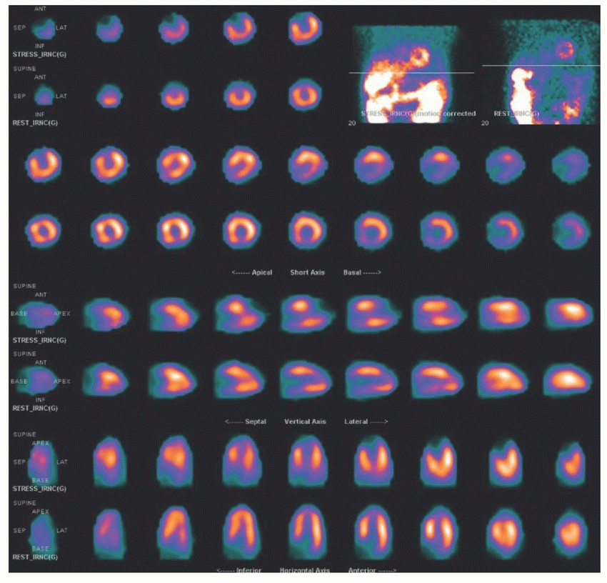
7a A 68-year-old lady underwent rest-stress Tc-99m tetrofosmin myocardial perfusion imaging for chest pain. What is the most likely diagnosis?
 |
A. Normal
B. Ischemia
C. Infarct
D. Attenuation artifact
View Answer
7a Answer B. The images demonstrate a large, severe intensity perfusion defect along the anteroseptal wall extending into the apex, which is not present (reversible) on resting images. Findings are most consistent with myocardial ischemia. A myocardial infarct would appear the same on stress and rest perfusion images. Although the breast attenuation artifact would be seen along the anteroseptal wall, it would present as a mild to moderate intensity fixed perfusion defect and not as a severe intensity reversible defect. Also, the cine images would show significant attenuation related to the breasts. Diaphragm attenuation artifact would be most prominent along the basal inferior wall.
References: Mettler FA, Guiberteau MJ. Essentials of nuclear medicine imaging, 6th ed. Philadelphia, PA: Saunders, 2012:145-151.
Ziessman HA, O’Malley JP, Thrall JH. Nuclear medicine: the requisites, 4th ed. Philadelphia, PA: Saunders, 2014:382-384, 395-400.
7b This territory is supplied by which of the following artery?
A. Left anterior descending artery (LAD)
B. Left circumflex artery (LCX)
C. Right coronary artery (RCA)
D. Ramus intermedius artery
View Answer
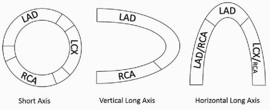
7b Answer A. Anterior, anteroseptal, and apical walls are typically supplied by the left anterior descending coronary artery (LAD). The left circumflex artery (LCX) typically supplies the lateral and inferolateral walls, while the inferior and inferoseptal walls are usually supplied by the right coronary artery (RCA) as shown in the image below.
 |
References: Mettler FA, Guiberteau MJ. Essentials of nuclear medicine imaging, 6th ed. Philadelphia, PA: Saunders, 2012:145-151.
Ziessman HA, O’Malley JP, Thrall JH. Nuclear medicine: the requisites, 4th ed. Philadelphia, PA: Saunders, 2014:382-384, 395-400.
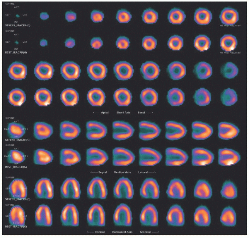
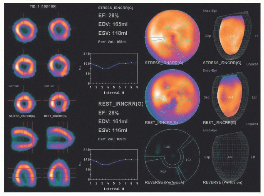
8 A 36-year-old man presents with shortness of breath. Based on the supplied resting and poststress myocardial perfusion SPECT images as well as the corresponding ejection fraction images, what is the most likely diagnosis?
 |
 |
A. Multivessel coronary artery disease
B. Right ventricular hypertrophy
C. Ischemic cardiomyopathy
D. Nonischemic cardiomyopathy
E. Subendocardial ischemia
View Answer
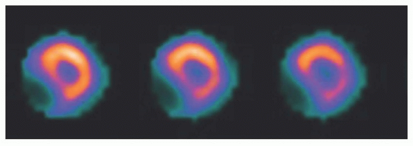
8 Answer D. Nonischemic cardiomyopathy frequently presents with normal perfusion but moderate to severe reduction in the ejection fraction. Often, heterogeneous perfusion without regional perfusion abnormalities may be seen. In contrast, ischemic cardiomyopathy is typically accompanied by a significant perfusion abnormality (ischemia or infarct) in a vascular territory. Right ventricular hypertrophy would result in straightening of the intraventricular septum, which is often referred to as the “D-shaped” septum (shown below).
 |
Reference: Danias PG, Ahlberg AW, Clark BA, III, et al. Combined assessment of myocardial perfusion and left ventricular function with exercise technetium-99m sestamibi gated single-photon emission computed tomography can differentiate between ischemic and nonischemic dilated cardiomyopathy. Am J Cardiol 1998;82(10):1253-1258.
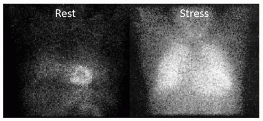
9 The following representative rest and poststress single frames from raw data cine images are from a patient who underwent thallium-201 exercise stress test. What is your interpretation?
 |
A. Bilateral pneumonia
B. Pulmonary sarcoidosis
C. Stress images acquired too early
D. High-risk coronary artery disease
E. Pulmonary hypertension
View Answer
9 Answer D. Increased Tl-201 uptake in the lungs, often quantified by increased lung to heart ratio, is associated with a higher incidence of more severe and multivessel coronary artery disease. Normal upper limit of lung to heart ratios has been reported to be in the range of 0.33 to 0.37. Lung to heart ratio of >0.45 correlates with a very high risk of severe, multivessel disease. It is thought to be the result of transient pulmonary edema due to stress-induced left ventricular dysfunction from myocardial ischemia.
References: Boucher CA, Zir LM, Beller GA, et al. Increased lung uptake of thallium-201 during exercise myocardial imaging: clinical, hemodynamic and angiographic implications in patients with coronary artery disease. Am J Cardiol 1980;46(2):189-196.
Kurata C, Tawarahara K, Taguchi T, et al. Lung thallium-201 uptake during exercise emission computed tomography. J Nucl Med 1991;32(3):417-423.
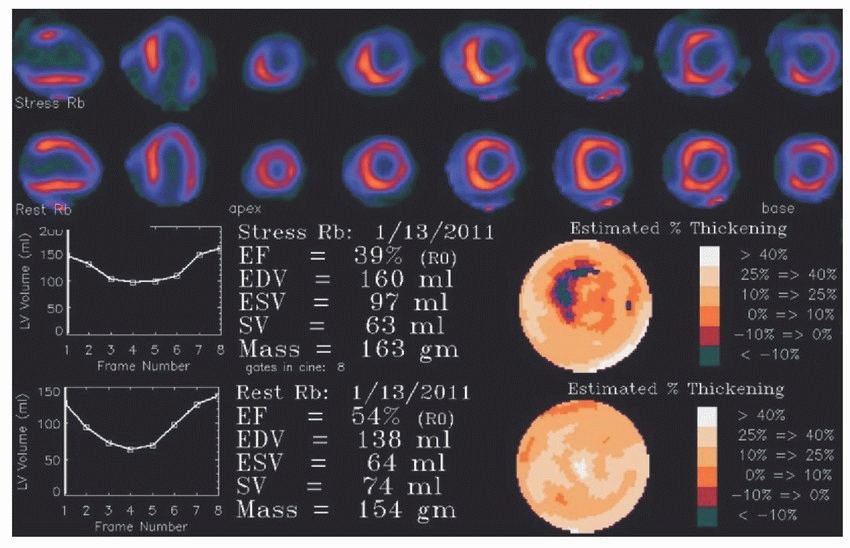
10 The following patient underwent a regadenoson pharmacologic stress test using Rb-82 rest-stress PET myocardial perfusion imaging. What is the most likely diagnosis?
 |
A. Left anterior descending artery disease
B. Left circumflex artery disease
C. Right coronary artery disease
D. Multivessel disease
View Answer
10 Answer D. The rest-stress perfusion images demonstrate a large severe intensity reversible perfusion abnormality along the anterior wall extending into the anterolateral and lateral walls. Also, there is evidence of stress-induced dilatation of the left ventricular (LV) cavity and stress-induced reduction of LV ejection fraction (LVEF) from 54% to 39%. Both of which suggest multivessel disease. Because of the short half-lives of current PET radiopharmaceuticals, the patient must undergo the stress perfusion PET imaging during the pharmacologic stress. As such, any reduction in the stress EF with PET represents myocardial stunning from significant ischemia with resultant decrease in myocardial contractility. On the other hand, with Tc-99m tetrofosmin and sestamibi, the patients usually wait 10 to 40 minutes between the injection at peak stress and imaging to allow for clearance from the liver. By this time, the stunned myocardium may have recovered and stress-induced reduction in the LVEF may have resolved. Transient ischemic dilatation (TID) is dilatation of the LV cavity on stress images compared to the rest images. It is secondary to combination of subendocardial ischemia and myocardial stunning.
References: Mettler FA, Guiberteau MJ. Essentials of nuclear medicine imaging, 6th ed. Philadelphia, PA: Saunders, 2012:170,172
Ziessman HA, O’Malley JP, Thrall JH. Nuclear medicine: the requisites, 4th ed. Philadelphia, PA: Saunders, 2014:382-384, 395-400.
11a A 63-year-old gentleman with ischemic cardiomyopathy is being evaluated for possible coronary artery bypass grafting with the hope of symptom improvement. Based on the following immediate (top row) and 24-hour delayed (bottom row) short-axis thallium-201 perfusion images, what is the MOST likely diagnosis?
 |
A. Ischemia
B. Attenuation artifact
C. Stunned myocardium
D. Hibernating myocardium
E. Nonviable myocardial infarct
View Answer
11a Answer E. The immediate images (top row) demonstrate a large-sized severe intensity perfusion defect along the inferolateral wall, which persists on 24-hour images (bottom row). Findings are consistent with a nonviable myocardial scar. Because of the redistribution phenomenon, Tl-201 would be expected to redistribute from the normal myocardium and localize in the chronically ischemic but viable (hibernating) myocardium on the delayed 6- or 24-hour images when compared to the immediate images. When identified, revascularization of the chronically ischemic but viable myocardium results in significant improvement in the myocardial function. Rest/redistribution Tl-201 myocardial scintigraphy or PET myocardial perfusion/F-18 FDG metabolism study may be used for the evaluation of myocardial viability.
References: Maddahi J, Schelbert H, Brunken R, et al. Role of thallium-201 and PET imaging in evaluation of myocardial viability and management of patients with coronary artery disease and left ventricular dysfunction. J Nucl Med 1994;35(4):707-715.
Ziessman HA, O’Malley JP, Thrall JH. Nuclear medicine: the requisites, 4th ed. Philadelphia, PA: Saunders, 2014:401-405.
11b What artery supplies this territory?
A. Left anterior descending artery
B. Left circumflex artery
C. Right coronary artery
D. Ramus intermedius artery
View Answer
11b Answer B. This area is typically supplied by the left circumflex artery as discussed in explanation 7b.
References: Mettler FA, Guiberteau MJ. Essentials of nuclear medicine imaging, 6th ed. Philadelphia, PA: Saunders, 2012:145-151.
Ziessman HA, O’Malley JP, Thrall JH. Nuclear medicine: the requisites, 4th ed. Philadelphia, PA: Saunders, 2014:382-384, 395-400.
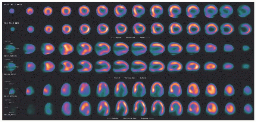
12 The patient is a 59-year-old gentleman with ischemic cardiomyopathy with LVEF of 35%. Based on the following resting N-13 ammonia (top row) and F-18 FDG- (bottom row) PET images, what is the most likely diagnosis?
 |
A. Ischemia
B. Attenuation artifact
C. Stunned myocardium
D. Hibernating myocardium
E. Nonviable myocardial infarct
View Answer


12 Answer D. The resting N-13 ammonia perfusion images demonstrate a large area of severe intensity perfusion defect along the inferolateral wall. Majority of this defect demonstrates metabolism on the F-18 FDG images. As such, the findings are consistent with a large zone of hibernating myocardium in the left circumflex artery territory. Hibernating myocardium is a chronically ischemic but viable myocardium, which should recover function after revascularization.
The patient undergoing F-18 FDG myocardial viability imaging undergoes glucose loading (sometimes with insulin) prior to F-18 FDG administration. This will help switch myocardial metabolism from fatty acids to glucose and increase the amount of F-18 FDG taken up in the heart. F-18 FDG-PET viability images should always be interpreted in conjunction with perfusion images. The perfusion defect is the only area that should be evaluated on the metabolic viability images as the normally perfused myocardium may utilize fatty acids and may not demonstrate significant FDG uptake. Hibernating myocardium will preferentially use glucose over free fatty acids and will demonstrate an area of increased FDG uptake. If this area corresponds to the area of resting perfusion abnormality, then it represents a hibernating myocardium. If the defect is present on both perfusion and metabolism images, then it likely represents a nonviable myocardial scar and the patient will not benefit from revascularization.
Stay updated, free articles. Join our Telegram channel

Full access? Get Clinical Tree


