| SKULL BASE REGION | Olfactory Groove |
| HISTOPATHOLOGY | Meningioma |
| PRIOR SURGICAL RESECTION | No |
| PERTINENT LABORATORY FINDINGS | N/A |
Case description
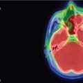
The patient is a 65-year-old man who was found to have an olfactory groove meningioma during workup for nasal polyps ( Figure 2.7.1 ). Serial neuroimaging showed a progressive increase in size with minimal surrounding edema. He had no significant symptoms, and olfaction was intact. Given the size, age, and medical comorbidities, the patient opted for radiosurgery. He underwent radiosurgery with 8 isocenters to cover a tumor volume of 6100 cc (margin dose 16 Gy; maximum dose 35.6 Gy; treated at 45% isodose line) ( Figure 2.7.2 ).
| Radiosurgery Machine | Gamma Knife – Perfexion |
| Radiosurgery Dose (Gy) | 16, at 45% isodose line |
| Number of Fractions | 1 |

Initial MRI demonstrating olfactory groove meningioma.

Imaging of the treatment plan.
| Critical Structure | Dose Tolerance |
|---|---|
| Optic nerve/chiasm |
|
| Pituitary gland | Stalk-to-gland radiation dose <0.8 |
| Side Effects/Complications | Frequency |
|---|---|
| Visual dysfunction | <1% with limited dose |
| Olfactory dysfunction | 34% change in smell with >5.1 Gy |
| Symptomatic edema | 5%–43% |
| Success Rate/Control Rate | Frequency |
|---|---|
| Progression-free survival | 97% at 5 years, 94.4% at 10 years |
| Local control | 71%–100% at 10 years |
Stay updated, free articles. Join our Telegram channel

Full access? Get Clinical Tree


