KEY FACTS
Terminology
- •
Membrane-covered midline abdominal wall defect with herniation of abdominal contents into base of cord
Imaging
- •
Liver + small bowel is most common type (large defects)
- •
Bowel-only type is smaller and more likely missed
- •
Omphalocele membrane is peritoneum + amnion
- ○
Mostly thin membrane but can also be cystic
- ○
- •
Umbilical cord inserts onto membrane (not always central)
- ○
Color Doppler best to show cord insertion site
- ○
- •
Ascites is common
- •
Membrane rupture is complication
Top Differential Diagnoses
- •
Normal physiologic bowel herniation
- ○
Bowel returns by 12 weeks
- ○
- •
Gastroschisis
- ○
Defect to right of normal cord insertion
- ○
Clinical Issues
- •
25-30% with associated anomalies
- ○
Cardiac defects common
- ○
- •
Chromosomal abnormalities in 30-40%
- ○
Bowel-only omphalocele with highest risk
- ○
- •
Syndromes associated with omphalocele
- ○
Beckwith-Wiedemann syndrome: Big tongue, macrosomia, large &/or horseshoe kidneys
- ○
Pentalogy of Cantrell: Part of heart in omphalocele sac
- ○
OEIS complex: O mphalocele, bladder e xtrophy, i mperforate anus, s pine anomaly
- ○
- •
Survival as high as 80-90% if normal chromosomes, no syndromes, and no other significant anomalies
Scanning Tips
- •
Evaluate abdominal wall cord insertion site at time of nuchal translucency screening
- •
Consider sac rupture if ascites suddenly resolves
- •
Consider formal echocardiogram in all cases
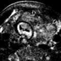
 and liver
  is covered by a membrane. The umbilical cord inserts directly onto the sac
  .
.
 and liver
  are seen well because of the presence of ascites
  . Also, there are some septations/cysts associated with the membrane
  , not uncommon since the membrane is peritoneum and amnion and can be thickened or cystic.
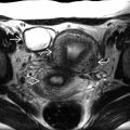
 , and umbilical cord insertion upon the membrane
  .
.
Stay updated, free articles. Join our Telegram channel

Full access? Get Clinical Tree


