Open Book Pelvis with Arterial Extravasation
Cody J. Schwartz
Daniel B. Nissman
CLINICAL HISTORY
45-year-old male with history of motor vehicle collision with tree, prolonged extrication, and hypotension en route to ED.
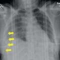
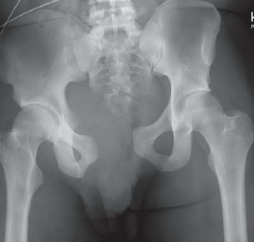 FIGURE 4A |
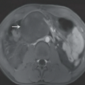
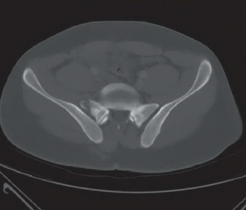 FIGURE 4B |
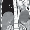
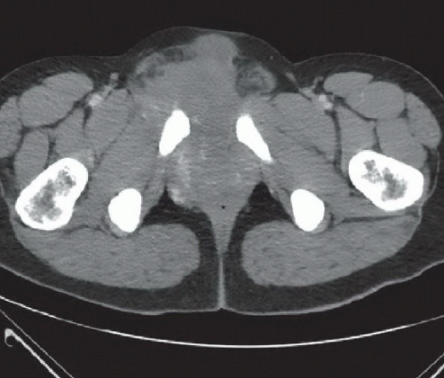 FIGURE 4C |
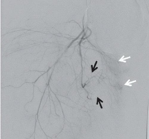 FIGURE 4D |
FINDINGS
Anteroposterior supine radiograph of the pelvis (Fig. 4A) demonstrates marked pubic symphysis diastasis to nearly 5 cm with marked right sacroiliac joint diastasis; likely right superior sacral ala fracture also noted. Axial CT image with bone window/level setting at the level of the superior right sacral ala (Fig. 4B) reveals a vertical sacral ala fracture. Axial CT image with soft tissue window/level setting (Fig. 4C) shows a large pelvic hematoma with areas of high density consistent with active extravasation. Selective right anterior iliac artery catheterization with angiogram (Fig. 4D) reveals contrast blush associated with multiple distal branches (white arrows); additional contrast blush was noted a few frames later nearby (black arrows).
DIFFERENTIAL DIAGNOSIS
Acetabulum fracture, pubic rami fractures, sacroiliac joint dislocation, open book pelvic fracture.
DIAGNOSIS
Related posts:


Stay updated, free articles. Join our Telegram channel

Full access? Get Clinical Tree


