Other Deep Gray Nuclei
Lubdha M. Shah, MD
IMAGING ANATOMY
Overview
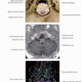
Red nucleus (RN)
Caudoventral magnocellular part (mcRN) and rostrodorsal parvocellular part (pcRN); latter represents majority of nuclear volume
Afferents from cerebral cortex on their distal dendrites and from deep cerebellar nuclei on their proximal dendrites and perikarya
mcRN mainly connected to motor and premotor cortices (arm and leg areas) and cerebellar interposed nuclei
Gives rise to crossed rubrospinal tract to spinal motor neurons and interneurons of distal, particularly flexor, muscles
Shows movement-related activity that correlates with duration, amplitude, and velocity of independent distal movements
pcRN receives afferents from primary, frontal, and supplementary motor cortices, accounting for 90% of total corticorubral afferents arising from cortical sublamina V
Projects onto bulbar olivary complex via central tegmental tract
Main efferent is represented by the inferior olive
Receives fibers from parts of cingulate and parietal cortices
May be involved in complex motor coordination rather than simple movements or may even be involved in nonmotor functions
Basal forebrain consists of several paleopallial structures, including substantia innominata, diagonal gyrus, and paraterminal gyrus
Substantia innominata and diagonal gyrus located in posterior 1/2 of anterior perforated substance
Paraterminal gyrus is on medial aspect of diagonal gyrus and posterior to subcallosal area
Nucleus basalis of Meynert (NB)
Within substantia innominata
Large cholinergic neurons
Projections from NB to cerebral cortex
Nucleus of the diagonal band of Broca in diagonal gyrus
Medial and lateral septal nuclei in paraterminal gyrus
Cholinergic projections from nucleus of diagonal band of Broca and septal nuclei to hippocampal formation
Locus coeruleus (LC)
Located adjacent to floor of 4th ventricle in rostral pons and extends into midbrain to level of inferior colliculi
LC cells are most densely populated at level of trochlear nucleus
Largest group of noradrenergic neurones in central nervous system
Excitatory projections to
Majority of cerebral cortex
Cholinergic neurons of basal forebrain
Cortically projecting neurons of thalamus
Serotoninergic neurons of dorsal raphe
Cholinergic neurons of pedunculopontine and laterodorsal tegmental nucleus
Inhibitory projections to sleep-promoting GABAergic neurons of basal forebrain and ventrolateral preoptic area
Implicated in modulating attentional states
Regulation of arousal and autonomic activity
Direct projections to spinal cord
Projections to autonomic nuclei: Dorsal motor nucleus of vagus, nucleus ambiguus, rostroventrolateral medulla, Edinger-Westphal nucleus, caudal raphe, salivatory nuclei, paraventricular nucleus, amygdala
LC activation produces increase in sympathetic activity and decrease in parasympathetic activity via these projections
Substantia nigra (SN): Pars compacta (SNPc) and pars reticulata (SNPr)
Part of A9 dopaminergic (DA) system
A9 DA fibers vary in size (20-40 µm) and extend from medial lemniscus to lateral cerebral peduncles
Sensorimotor areas of striatum project to ventrolateral pallidum and ventrolateral SNPc cell columns
Projections from central striatum terminate more centrally in both pallidum and ventral densocellular SNPc
Ventral striatum projects topographically into ventral pallidum, ventral tegmental area, and densocellular SNPc
Pars compacta produce dopamine → increased motor activity
Ventral tegmental area (VTA)
Medial to substantia nigra in rostral mesencephalon
Heterogeneous groups of neurons, part of A10 DA system
A10 DA fibers within ventromedial midbrain, consist of small diameter (15-30 µm), non-myelinated axons that ascend in medial forebrain bundle
Synthesizes dopamine, which is sent to nucleus accumbens (NA)
Reciprocal connections with limbic cortices through mesolimbic pathway
Including NA, amygdala, cingulate cortex, and hippocampal complex
Stay updated, free articles. Join our Telegram channel

Full access? Get Clinical Tree


