Parahippocampal Gyrus (Areas 28, 34, 35, 36)
Jared A. Nielsen, PhD
Jeffrey S. Anderson, MD, PhD
Key Facts
Location and Boundaries
Areas 28, 34, 35, 36: Located on parahippocampal gyrus, extending lateral and posterior from hippocampus
Entorhinal cortex (areas 28, 34): Comprises anteromedial parahippocampal gyrus
Perirhinal cortex (area 35): Immediately lateral to entorhinal cortex
Entorhinal cortex separated from perirhinal cortex by rhinal (collateral) sulcus
Ectorhinal cortex (area 36): Posterolateral to perirhinal cortex
Function
Spatial navigation: Map of spatial location and trajectory in entorhinal cortex with grid cells encoding spatial location and path cells encoding direction
Olfaction: Primary olfactory cortex located at anterior margin of entorhinal cortex at level of anterior margin of amygdala
Encoding visual scenes: Parahippocampal place area (PPA) in posterior parahippocampal gyrus active during perception of scenes (e.g., buildings, houses, locations)
Memory: Bridge between hippocampus and neocortex for memory encoding and retrieval
Area 28-, 34-36-Associated Disorders
Alzheimer dementia: Entorhinal cortex shows greatest atrophy, site of earliest pathologic changes
Medial temporal sclerosis: Variable involvement of hippocampus, amygdala, and entorhinal/perirhinal cortex
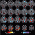
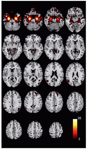 (Left) Coactivation map of Brodmann areas 28, 34, 35, and 36 shows brain regions that reliably activate with the centroid of voxels lying within areas 28, 34, 35, and 36 in over 4,000 studies from the NeuroSynth database. Image is the average of left and right coactivation maps. |
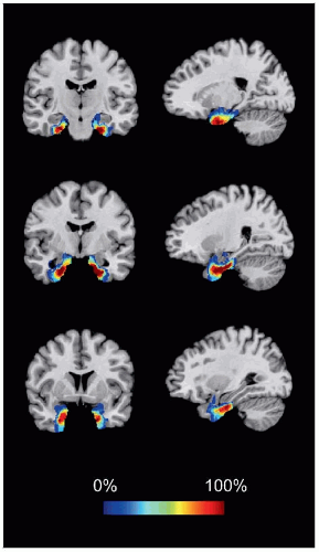 (Right) Coronal and axial slices from a cytoarchitectonic map of parahippocampal gyrus is shown. This quantitative probabilistic map was derived from postmortem human brains and is specific to cellular properties unique to areas 28, 34, 35, and 36 (data source: SPM Anatomy toolbox). |
LOCATION AND BOUNDARIES
Location
Areas 28, 34, 35, and 36: Parahippocampal gyrus, extending lateral and posterior from hippocampus
Area 28 (ventral entorhinal) and area 34 (dorsal entorhinal): Adjacent to hippocampal subiculum
Area 35 (perirhinal cortex) and area 36 (ectorhinal, parahippocampal cortex): Occupy lateral parahippocampal and anterior fusiform gyri
Boundaries
Entorhinal cortex (areas 28, 34): Comprises anteromedial parahippocampal gyrus
Separated from hippocampus by hippocampal fissure
Boundary with subiculum at medial margin ventral to hippocampal fissure
Separated from perirhinal cortex by collateral sulcus
Areas 28 and 34 separated by tentorial notch
Posteriorly contiguous with retrosplenial cingulate
Perirhinal cortex (area 35): Immediately lateral to entorhinal cortex
Includes medial bank of collateral sulcus
Bordered by temporal pole (area 38) rostrally
Anterior margin 2-3 mm anterior to limen insulae at anterior margin of collateral sulcus (˜ 24 mm caudal to temporal pole)
Ectorhinal cortex (area 36): Caudal to perirhinal cortex
Overlapping terms: Ectorhinal, postrhinal, parahippocampal cortex
Includes anterior medial fusiform gyrus
Lateral margin at occipitotemporal sulcus, separating perirhinal from inferior temporal cortex (area 20)
Bordered by fusiform gyrus (area 37) caudally
FUNCTION
Related posts:
Stay updated, free articles. Join our Telegram channel

Full access? Get Clinical Tree


