Passive Reading
Lubdha M. Shah, MD
Key Facts
Anatomy-based Imaging Issues
Primarily for localization of language comprehension areas in dominant hemisphere region of posterior superior temporal gyrus/parietal angular gyrus (Wernicke area)
Usually yields an additional language response in region of dominant hemisphere lateral inferior/middle frontal gyri (Broca area)
Language processing
Meaning of words and sentences (semantics)
Sound structure of words (phonology)
Rules for combinations of words (syntax)
Reading (and passive viewing) involves automatic semantic processing
Design
Block design: 4 minutes
Presentation of text reading blocks alternating with blank screen fixation
Visual presentation of short (2-4 sentences) narrative paragraphs for 10 seconds each
2 consecutive narrative text presentations per 20-second block
Comparison can be made to activation produced by nonsense reading
Video feed can evaluate whether patient’s eyes are open
Ask subject after the scan if he/she was able to keep eyes focused on center of screen
Applications
Presurgical planning for language lateralization
Particularly in patients with temporal lesions
Combined task analysis of several language tasks is more robust and reliable compared to individual task analysis
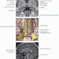
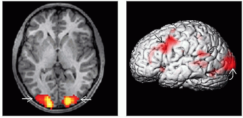 (Left) Axial MPRAGE with overlay of BOLD signal during reading task illustrates expected activation in bilateral visual cortices in the occipital lobes  . (Right) 3D surface rendering displays activation in the receptive speech area . (Right) 3D surface rendering displays activation in the receptive speech area  . Activation in the premotor . Activation in the premotor  and visual and visual  areas is also noted. Anterior temporal cortex is involved in semantic and syntax processes whereas the posterior temporal lobe is activated during language comprehension. areas is also noted. Anterior temporal cortex is involved in semantic and syntax processes whereas the posterior temporal lobe is activated during language comprehension. |
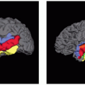
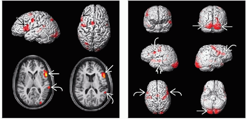 (Left) 3D surface rendering images (top) demonstrate left IFG activation
 during a reading task. Axial MPRAGE with BOLD overlay (bottom) show this robust expressive speech activation during a reading task. Axial MPRAGE with BOLD overlay (bottom) show this robust expressive speech activation  and activation in the receptive speech area and activation in the receptive speech area  . (Right) 3D surface rendering images during a passive reading task show activation in bilateral IFG . (Right) 3D surface rendering images during a passive reading task show activation in bilateral IFG  in this patient with bilateral expressive speech representation and mild activation in the left receptive speech area in this patient with bilateral expressive speech representation and mild activation in the left receptive speech area  . Expected activation is noted in bilateral occipital visual cortices . Expected activation is noted in bilateral occipital visual cortices  . .Related posts:Stay updated, free articles. Join our Telegram channel
Full access? Get Clinical Tree
 Get Clinical Tree app for offline access

|