Pediatric Nuclear Medicine
QUESTIONS
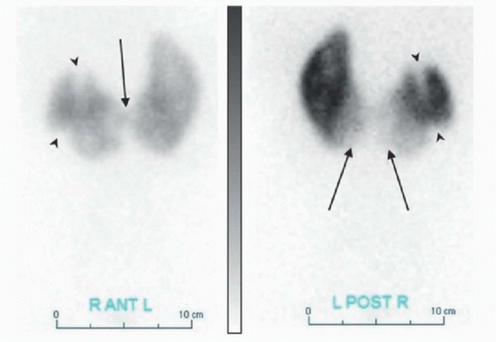
1A The following renal scintigraphy was performed on a 7-year-old patient with fever. What radiopharmaceutical was used for this examination?
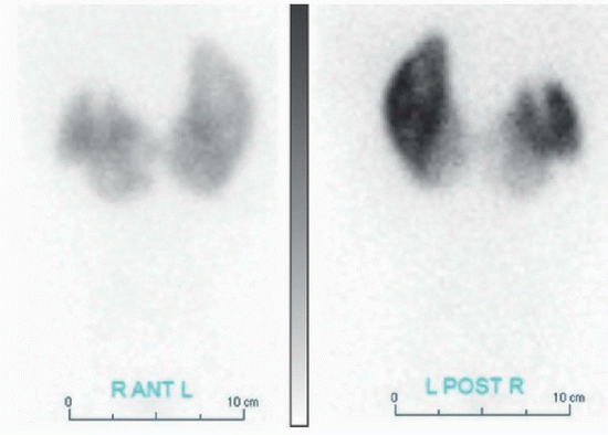 |
A. Tc-99m DMSA
B. Tc-99m DTPA
C. Tc-99m MAG3
D. I-123 hippuran
View Answer
1A Answer A. Tc-99m DMSA (dimercaptosuccinic acid) is commonly used to detect pyelonephritis and renal scarring due to vesicoureteral reflux in the pediatric population. Approximately 40% of the injected radiopharmaceutical binds to the sulfhydryl groups of the proximal renal tubules and remains there, thus allowing for high-count renal cortical imaging. MAG3 (mercaptoacetyltriglycine) is a tubular-secreting agent; it is the most commonly used radiopharmaceutical for the evaluation of the renal flow and function. DTPA (diethylenetriaminepentaacetic acid) is a glomerular filtration agent, which can be used to calculate the glomerular filtration rate (GFR); while DTPA can also be used to examine the renal flow and function, it has poor image quality compared to MAG3 due to significantly higher background activity and slower renal clearance. I-123 hippuran undergoes tubular secretion and glomerular filtration allowing for assessment of renal function. It has been replaced by MAG3.
References: Mettler FA, Guiberteau MJ. Essentials of nuclear medicine imaging, 6th ed. Philadelphia, PA: Saunders, 2012:316, 321, 336-338.
Ziessman HA, O’Malley JP, Thrall JH. Nuclear medicine: the requisites, 4th ed. Philadelphia, PA: Saunders, 2014:173-174, 198-200.
1B What is the most likely diagnosis?
A. Severe renal dysfunction
B. Bilateral hydronephrosis
C. Pyelonephritis
D. Wilms tumor
E. Lymphoma
View Answer
1B Answer C. Anterior and posterior Tc-99m DMSA planar images demonstrate abnormal renal axis with tissue joining the lower poles of each kidney (arrow) consistent with a horseshoe kidney. Additionally, multiple areas of decreased radiotracer uptake (arrowheads) are identified within the upper and lower pole of the right kidney. In the setting of acute febrile illness, findings are most compatible with pyelonephritis.
 |
References: Mettler FA, Guiberteau MJ. Essentials of nuclear medicine imaging, 6th ed. Philadelphia, PA: Saunders, 2012:316, 321, 336-338.
Ziessman HA, O’Malley JP, Thrall JH. Nuclear medicine: the requisites, 4th ed. Philadelphia, PA: Saunders, 2014:295, 297.
2 What is an advantage of the contrast voiding cystourethrogram (VCUG) compared to the direct radionuclide cystogram (RNC)?
A. Lower radiation
B. Better anatomic detail
C. Greater sensitivity
D. Bladder volume calculation
View Answer
2 Answer B. The main advantages of contrast voiding cystourethrogram (VCUG) are superior anatomic detail and more accurate grading of the vesicoureteral reflux (VUR). VCUG is recommended for the initial workup of boys with vesicoureteral reflux to exclude an anatomic cause of the reflux (i.e., posterior urethral valve). Advantages of direct radionuclide cystography (RNC) include greater sensitivity for the detection of reflux, lower radiation, and the ability to calculate the residual bladder volume. RNC allows for continuous imaging of the urinary tract throughout the examination and thus may be more sensitive in the diagnosis of significant VUR. However, due to low anatomic resolution, it is more likely to miss a low-grade reflux. While the use of modern grid-controlled variable-rate pulsed fluoroscopy reduces the effective radiation dose by about eightfold compared to the older continuous fluoroscopy units, effective dose of VCUG performed with this technique is still about ninefold higher than that received during RNC.
References: Mettler FA, Guiberteau MJ. Essentials of nuclear medicine imaging, 6th ed. Philadelphia, PA: Saunders, 2012:338.
Ward VL, Strauss K, Barnewolt C, et al. Pediatric radiation exposure and effective dose reduction during voiding cystourethrography. Radiology 2008;249(3):1002-1009.
Ziessman HA, O’Malley JP, Thrall JH. Nuclear medicine: the requisites, 4th ed. Philadelphia, PA: Saunders, 2014:201-203.
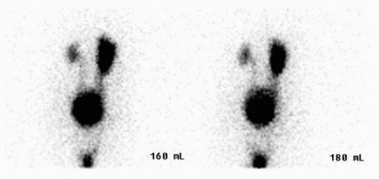
A. Mild left, moderate right
B. Moderate left, mild right
C. Severe left, moderate right
D. Moderate left, severe right
E. Mild bilateral
F. Moderate bilateral
G. Severe bilateral
View Answer
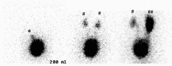
3 Answer D. Posterior images acquired during the direct radionuclide cystography (RNC) demonstrate reflux of radiopharmaceutical into bilateral renal pelvicalyceal systems. There is dilatation of the right collecting system. Findings represent moderate left-sided and severe right-sided vesicoureteral reflux (VUR). Direct RNC with bladder catheterization is the most commonly used technique for diagnosing reflux. Dynamic posterior imaging is acquired during the bladder filling, voiding, and after voiding. Grading of VUR is as follows: mild—visualization of the ureters (* in the image below); moderate—visualization of the pelvicalyceal system (# in the image below); and severe—visualization of pelvicalyceal system with dilated collecting system (## in the image below). Grading is important as renal damage is more likely in severe VUR compared to mild or moderate grades. RNC is the technique of choice for the evaluation and follow-up of the children with UTIs and suspected vesicoureteral reflux. It is also used in screening the siblings of the patients with VUR. Tc-99m sulfur colloid and Tc-99m DTPA are the radiopharmaceuticals most commonly used as they do not get absorbed by the epithelial lining of the bladder or ureters.
 |
References: Mettler FA, Guiberteau MJ. Essentials of nuclear medicine imaging, 6th ed. Philadelphia, PA: Saunders, 2012:338.
Sandler M. Diagnostic nuclear medicine, 4th ed. Philadelphia, PA: Lippincott Williams & Wilkins, 2003:1100-1102.
Ziessman HA, O’Malley JP, Thrall JH. Nuclear medicine: the requisites, 4th ed. Philadelphia, PA: Saunders, 2014:201-203.
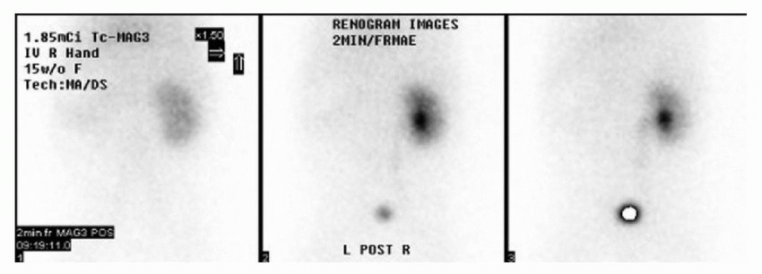
4 A 4-month-old child has a left kidney containing multiple noncommunicating cysts with abnormal echogenic renal parenchyma on ultrasound. When read in conjunction with this renal scintigraphy, what is the most likely diagnosis?
 |
A. Autosomal recessive polycystic kidney disease
B. Congenital mesoblastic nephroma
C. Ureteropelvic junction obstruction
D. Multicystic dysplastic kidney
View Answer
4 Answer D. The classic appearance of multicystic dysplastic kidney (MCDK) on ultrasound is that of a kidney containing multiple noncommunicating cysts of various sizes without normal parenchyma. When evaluated by renal scintigraphy, an MCDK will lack normal functioning renal parenchyma, which can confirm the diagnosis. The remaining entities will have some functioning renal tissue. Polycystic kidney disease is a bilateral process and the right kidney is normal in this case.
References: Sandler M. Diagnostic nuclear medicine, 4th ed. Philadelphia, PA: Lippincott Williams & Wilkins, 2003:1091.
Coley B. Caffey’s pediatric diagnostic imaging, 12th ed. Philadelphia, PA: Elsevier Saunders, 2013:1195.
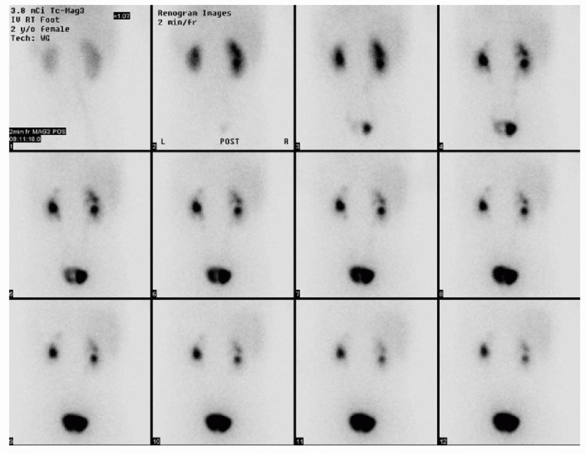
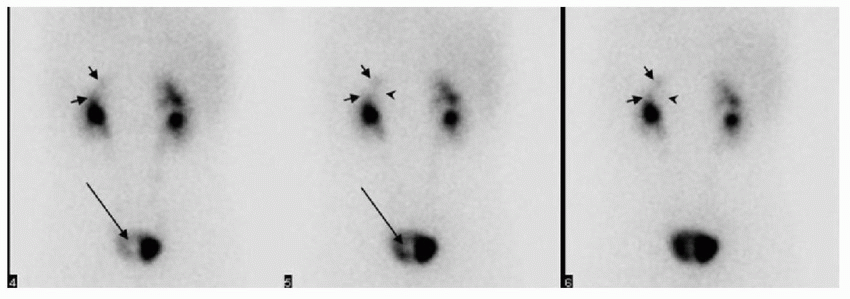
5 A 2-year-old girl has a known renal anomaly on ultrasound. Renal scintigraphy was performed to evaluate the renal function. What is the most likely diagnosis?
 |
A. Calculus
B. Diverticulum
C. Ureterocele
D. Rhabdomyosarcoma
View Answer
5 Answer C. The early images demonstrate a rounded area of photopenia within the left side of the bladder (long arrow), which is most compatible with a ureterocele. On more delayed images, a left upper pole moiety (short arrows) with central area of photopenia (arrowhead) is identified. This is most compatible with a hydronephrotic left upper pole moiety with a moderately impaired renal function. Normal functioning left lower pole moiety is seen without hydronephrosis. Duplex collecting system is also identified on the right side with normally functioning, nondilated upper and lower pole moieties.
Ectopic ureterocele is commonly found in the setting of a duplicated system and is almost always associated with the upper pole moiety. Lower pole moiety is typically associated with reflux. As the Weigert-Meyer rule states, the ureter from the upper pole moiety of a completely duplicated collecting system inserts inferior and medial to the normal insertion site of the lower pole moiety in the trigone. In patients with duplication, MAG3 renogram is typically done to assess the function of the obstructed upper pole moiety. Bladder calculus and rhabdomyosarcoma may also cause a photopenic defect in the bladder but are not associated with renal duplication. Diverticulum would appear as an intense outpouching from the bladder.
 |
References: Coley B. Caffey’s pediatric diagnostic imaging, 12th ed. Philadelphia, PA: Elsevier Saunders, 2013:1091.
Sandler M. Diagnostic nuclear medicine, 4th ed. Philadelphia, PA: Lippincott Williams & Wilkins, 2003:1200-1202.
6 An 18-month-old patient presents with a high clinical suspicion for child abuse and equivocal findings on the skeletal survey. What site of fracture has the highest specificity for the nonaccidental trauma on a bone scintigraphy?
A. Skull
B. Scapula
C. Clavicle
D. Long bone diaphysis
View Answer
6 Answer B. Shaking injury may result in scapular fractures, which have a high specificity for child abuse. Skull, diaphyseal long bone, and clavicle fractures have lower specificity. While skull fractures are seen on the radiographs, they may be difficult to see on the bone scan. Also, subtle fractures along the growth plates are best seen on radiographs owing to the intense physiologic physeal uptake on the bone scan. As such, skeletal survey using radiographs is more sensitive than bone scintigraphy. Bone scintigraphy is best utilized when the clinical suspicion for nonaccidental trauma is high but the skeletal survey is inconclusive.
References: Coley B. Caffey’s pediatric diagnostic imaging, 12th ed. Philadelphia, PA: Elsevier Saunders, 2013:1588.
Habibian MR. Nuclear medicine imaging: a teaching file, 2nd ed. Philadelphia, PA: Lippincott Williams & Wilkins, 2009:377-378.
7 A 15-year-old runner has three-phase bone scintigraphy performed for lower leg pain. What characteristic finding would establish the diagnosis of shin splints?
A. Three-phase positive
B. Fusiform shape
C. Posteromedial cortex
D. Focal cortical uptake
View Answer
7 Answer C. Characteristics of shin splints on the bone scan include linear cortical uptake along the posteromedial cortex of at least one-third of the tibial diaphysis on delayed imaging that is negative on the flow and blood pool phases. Fusiform shape, focal cortical uptake, and three-phase positive uptake are typical findings associated with a stress fracture.
References: Mettler FA, Guiberteau MJ. Essentials of nuclear medicine imaging, 6th ed. Philadelphia, PA: Saunders, 2012:294.
Ziessman HA, O’Malley JP, Thrall JH. Nuclear medicine: the requisites, 4th ed. Philadelphia, PA: Saunders, 2014:307-314.
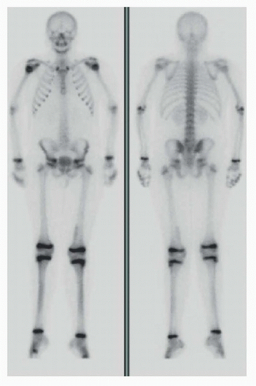
8 The following anterior and posterior bone scintigraphy images were acquired on a 19-year-old with low back pain. The nuclear medicine technologist asks you to review the images prior to the patient leaving the department. What is the most appropriate next step?
 |
A. MRI of the lumbar spine
B. CT of the lumbar spine
C. Radiographs of the lumbar spine
D. SPECT images of the lumbar spine
E. No further imaging necessary
View Answer
8 Answer D. Spondylolysis is a common cause of lower back pain in young adults, especially athletes. Repetitive trauma is thought to be the precipitating factor leading to stress reaction and eventual disruption of the par interarticularis. This may occur unilaterally or bilaterally and occurs most commonly at the L5 level. Bone scintigraphy is more sensitive than radiographs for localizing the site of bone stress, and in this patient, the initial radiographs were negative. At initial workup, both plain film of the lumbar spine and planar bone scintigraphy may be unrevealing. In these instances, SPECT or SPECT/CT imaging may show increased uptake at the par interarticularis. Additionally, SPECT can provide further anatomic localization allowing for the distinction of uptake at the pars abnormality or that at the facet joints. Furthermore, SPECT/CT will demonstrate the presence or absence of a pars defect and its associated changes, such as sclerosis. As seen with this case, a subtle abnormality on planar bone imaging is readily visible on the SPECT images (below), which revealed a focus of intense uptake at the left L5 level consistent with spondylolysis.
 |
References: Sandler M. Diagnostic nuclear medicine, 4th ed. Philadelphia, PA: Lippincott Williams & Wilkins, 2003:1112.
Ziessman HA, O’Malley JP, Thrall JH. Nuclear medicine: the requisites, 4th ed. Philadelphia, PA: Saunders, 2014:110.
Zukotynski K, Curtis C, Grant FD, et al. The value of SPECT in the detection of stress injury to the pars interarticularis in patients with low back pain. J Orthop Surg Res 2010;5:13.
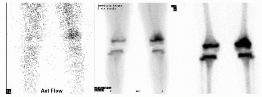
9 A 7-year-old male presents with left knee pain and fever for 2 days with negative radiographs. The patient was unable to undergo MR due to cochlear implants. Instead, three-phase bone scintigraphy was performed. What is the most likely diagnosis?
 |
A. Septic arthritis
B. Osteosarcoma
C. Fracture
D. Osteomyelitis
View Answer
9 Answer D. The supplied three-phase images acquired after the IV injection of diphosphonate demonstrate regional area of increased blood flow to the distal left femur, which further localizes to the distal femoral metaphysis on the blood pool and the delayed images. Findings represent osteomyelitis. While this is the most common appearance of osteomyelitis on bone scintigraphy, it is important to remember that early osteomyelitis in children may present as a cold or photopenic defect of the metaphysis with increased activity in the adjacent diaphysis; this is thought to result from increased intramedullary pressure or thrombosed vessels.
Osteomyelitis is a common pediatric problem and frequently involves the highly vascularized metaphysis of fast-growing bones. Distal femur and proximal tibia are the most common locations. Initial radiographs for osteomyelitis are often negative, especially in the first few weeks. MRI is regarded as the optimum imaging modality in the evaluation of infection. However, if there is concern for multifocal infection, bone scintigraphy can offer an advantage over MRI because whole-body images are routinely included with scintigraphy. If there are contraindications to MR imaging or if sedation may be required for the MRI, then bone scintigraphy should be considered since it offers greater sensitivity than radiograph and can detect changes as early as 24 hours.
References: Coley B. Caffey’s pediatric diagnostic imaging, 12th ed. Philadelphia, PA: Elsevier Saunders, 2013:1471-1474.
Sandler M. Diagnostic nuclear medicine, 4th ed. Philadelphia, PA: Lippincott Williams & Wilkins, 2003:1108.
Ziessman HA, O’Malley JP, Thrall JH. Nuclear medicine: the requisites, 4th ed. Philadelphia, PA: Saunders, 2014:123-124.
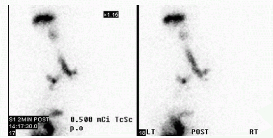
10 The shown examination was performed on a child with neurologic impairment. Which of the following is the most likely indication?
 |
A. Gastroesophageal reflux
B. Esophageal dysmotility
C. Gastric emptying
D. Aspiration
View Answer
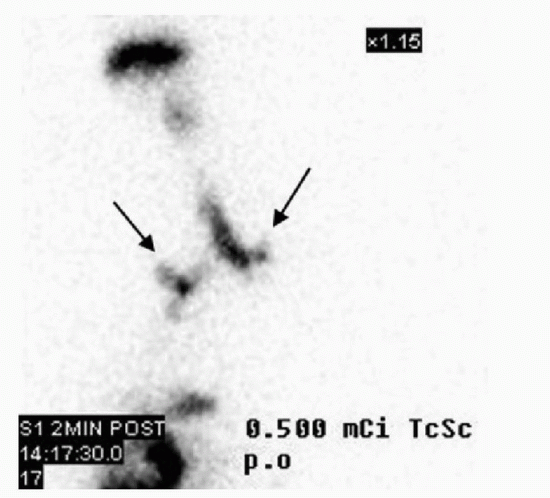
10 Answer D. A salivagram is a sensitive yet simple nuclear medicine study to evaluate for aspiration. At our institution, Tc-99m sulfur colloid (0.3 to 0.4 mCi) is mixed in water and placed on the back of the tongue with the patient in the supine position. Dynamic acquisition for 1 hour is performed followed by static images at 1 and 3 hours. This case is grossly positive with abrupt aspiration of the radiotracer into the tracheobronchial tree (arrows below). Gastroesophageal reflux, gastric emptying, and esophageal motility are not evaluated on the salivagram scintigraphy. This exam is more sensitive than reflux scintigraphy in detecting aspiration.
 |
Reference: Ziessman HA, O’Malley JP, Thrall JH. Nuclear medicine: the requisites, 4th ed. Philadelphia, PA: Saunders, 2014:288-294.
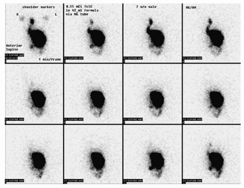
11A Which of the following statements is the most accurate regarding the findings in this 6-month-old infant?
 |
Stay updated, free articles. Join our Telegram channel

Full access? Get Clinical Tree


