Abstract
The treatment options for pyometra include antibiotic administration, cervical dilation with drainage, and surgical intervention. Surgical management options include laparotomy with lavage and/or total hysterectomy. However, reports on the percutaneous drainage of pyometra are rare. We present a case report and discussion of pyometra in a woman in her 70s with pyometra caused by cervical invasion of rectal cancer and fibrosis following radiation therapy. Antibiotic treatment was ineffective, and computed tomography (CT)-guided transabdominal uterine drainage was performed. After drainage, the abscess reduced in size, and no recurrence was observed during the subsequent 12 months. Percutaneous drainage should be considered as a treatment option in cases where antibiotics are ineffective, and transvaginal drainage is not feasible.
Introduction
Pyometra is characterized by the accumulation of purulent material within the uterine cavity due to obstruction of the cervical canal or vagina. Treatment options include antibiotic therapy, transvaginal drainage and surgical interventions such as laparotomy with irrigation. Laparotomy with abdominal lavage and/or total hysterectomy is often performed in cases of pyometra perforation.
Here, we report a case of perforated pyometra in which both abdominal lavage and transvaginal drainage were unfeasible owing to tumor invasion and fibrosis previously caused by radiation therapy. The patient underwent computed tomography (CT)-guided transabdominal uterine drainage, which resulted in a favorable clinical outcome.
Case report
A woman in her 70s, with a history of abdominoperineal resection for rectal cancer performed 17 years prior, presented at our hospital with worsening discomfort in the anorectal region. CT and magnetic resonance imaging (MRI) revealed lung metastases and local recurrence with cervical invasion. The patient was undergoing chemoradiotherapy; however, during radiotherapy, she developed fever and fatigue and was diagnosed with cervical invasion-induced pyometra. Despite antibiotic therapy, the patient’s symptoms persisted. The attempted transvaginal drainage procedure failed because of cervical obstruction caused by tumor invasion. The patient was then referred to our department for uterine drainage.
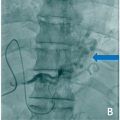
Physical examination revealed a temperature of 38.1°C, generalized fatigue, pain from the right buttock to the posterior thigh, sensory impairment in the right sole, and evidence of an inflammatory response (white blood cell count of 19,300 /mm³ and C-reactive protein of 13.54 mg/dL). Abdominal contrast-enhanced CT showed fluid accumulation within the uterine cavity with peripheral enhancement, suggestive of pyometra ( Fig. 1 ). Contrast-enhanced MRI revealed cervical and sacral invasions. Additionally, a rupture was observed in the posterior uterine wall with a fistula extending to the anterior sacrum. The pyometra content leaked into the anterior sacrum and spread to the left gluteus medius ( Fig. 2 ).


Related posts:
 A rare and life-threatening case of spontaneous hemopneumothorax presenting with severe hemodynamic instability
 Avascular necrosis of lateral femoral condyle in a middle-aged woman from central India
 A very rare case of ileocolic and appendiceal intussusception with acute appendicitis
 Hepatic and extra-hepatic hydatid cysts: A case series of radiological and clinical insights
 Percutaneous transhepatic embolization of gastro-esophageal varices for the treatment of variceal bleeding in portal vein thrombosis secondary to hepatocellular carcinoma: A case report
 Successful endovascular treatment for pediatric ruptured cervical spinal perimedullary arteriovenous fistula: A case report
Stay updated, free articles. Join our Telegram channel

Full access? Get Clinical Tree


