Abstract
Objective
Fat embolism syndrome and cerebral fat emboli are rare yet serious conditions arising from systemic distribution of bone marrow emboli. Emboli are known to produce high-intensity transient signals (HITS) in a Doppler signal. We hypothesized that both intramedullary nailing in pigs and median sternotomy in human infants cause bone marrow release, that some of these cause cerebral emboli, and that these were detectable by a new cerebral doppler ultrasound monitoring system (NeoDoppler). We also aimed to describe the intensity of HITS generated during these procedures.
Methods
Specific pathogen-free Norwegian landrace pigs were allocated to either bilateral femoral nailing or injection of autologous bone marrow (positive controls). Testing was carried out under continuous Doppler monitoring. Presence of cerebral emboli was confirmed with histology. NeoDoppler data from infants undergoing sternotomy prior to cardiac surgery were investigated for comparison.
Results
Eleven of twelve pigs were monitored with cerebral Doppler ultrasound during femoral surgery. HITS were seen in five (45%). Brain biopsies demonstrated bone marrow emboli in 11 of the 12 (92%). Four positive control pigs received intraarterial injections of bone marrow, saline, or contrast, and strong HITS were detected in all pigs (100%). Median sternotomy in eight human infants was associated with a significant increase in embolic burden; the HITS intensity was lower than HITS in pigs.
Conclusion
High-frequency cerebral Doppler ultrasound is a valuable tool for perioperative monitoring that can detect emboli in real-time, but sensitivity and specificity for bone marrow emboli may be limited and size-dependent.
Introduction
Fat embolism syndrome (FES) is a serious condition defined by the presence of fat globules in the systemic and pulmonary circulation [ ]. The syndrome is commonly seen in trauma patients with long bone fractures or after orthopedic procedures but has also been reported following procedures such as liposuction and bone marrow biopsy [ ]. Cardiac surgery, including sternotomy, in children is of special concern as cardiac shunts are common and may transport emboli directly into the systemic circulation [ ]. In rare cases, cerebral symptoms present in isolation, a condition referred to as cerebral fat embolism (CFE). CFE can be challenging to diagnose and may result in the delay of appropriate treatment [ ]; CFE may also occur in the absence of intracardiac shunt, such as patent foramen ovale [ ]. Although magnetic resonance imaging (MRI) is considered the gold standard for CFE diagnosis [ , ], it is uncertain if early MRI may prove diagnostic.
Cerebral Doppler ultrasound can provide real-time detection of emboli in the cerebral circulation and may thus be used to quantify embolic load during medical procedures [ ]. Solid or gaseous entities in the blood stream have higher reflectivity than surrounding erythrocytes and produce characteristic high-intensity transient signals (HITS) in the Doppler spectrum. Transcranial Doppler ultrasound has been used to detect HITS during various medical procedures [ ], including orthopedic surgery [ ] and cardiac surgery in infants [ ]. The clinical significance of the presence of HITS has, however, not yet been established and there is a lack of studies correlating HITS characteristics with histologically verified brain emboli.
In this study, we aim to describe the relationship between surgically generated bone marrow emboli and ultrasound-detected HITS in vivo . To this end, we performed bilateral reamed intramedullary nailing of the femur in pigs without intracardiac shunt. Timing, presence, and extent of CFE was assessed by combining cerebral Doppler ultrasound monitoring, postmortem MRI, and histological examination of brain tissue. For validation, bone marrow was injected in the carotid artery of four additional animals with identical monitoring. Data from infant cardiac surgery were used for comparison.
Materials and methods
Part 1: HITS monitoring during femoral nailing in a porcine model
Study animals
Twelve specific pathogen-free Norwegian landrace pigs were assessed for inclusion in March–September 2022 [ ]. Exclusion criteria were pre-existing disease, complications not caused by the femoral nailing itself, and patent foramen ovale . No animals were excluded prior to surgery.
Experimental procedure
A detailed protocol describing the surgery and anesthesia of this study has been published by Kristiansen et al. [ ]. Data were collected through multimodal monitoring and postmortem examinations ( Fig. 1 ). Biopsies were collected postmortem from lungs, heart, kidneys, and brain. The head was transferred to a nearby MRI facility and scanned within one hour of sacrifice.

Doppler ultrasound monitoring with the NeoDoppler system
The NeoDoppler ultrasound system was developed by the ultrasound group at NTNU and a research set-up was used in this study [ ]. The single element plane wave transducer operates at a frequency of 7.8 MHz. It is a multigated Doppler, which makes it possible to measure blood flow velocities in several depths simultaneously down to approximately 35 mm. All depths are recorded, and the depth of interest is selected during analysis. Anatomical structures cannot be visualized as the set-up cannot produce B mode images and no angle correction is performed. Thermal and mechanical indices are kept within the recommended limits for long-term continuous monitoring, as verified with calibrated hydrophone measurements. The NeoDoppler ultrasound system can measure rapid changes in blood flow [ ] and has been used to map the cerebral effects of general anesthesia [ ] and detect HITS, trends and events [ , ] during cardiac surgery in infants. Data processing was performed with an in-house software developed in MATLAB (MathWorks© R2017a).
Trepanning was used to create a fontanel-like acoustic window to the cerebral blood flow. An L-incision was made, starting from the base of the ear to the midline, and perpendicular along the midline in anterior direction. The skull bone was then uncovered, and a circular opening with diameter of 2–3 cm was drilled with a Dremel 4000 multitool with flexible shaft and 6.4/7.9 mm Dremel carving bits (Dremel Europe, Breda, Netherlands) without penetrating the dura. The NeoDoppler probe was then placed on the dura and positioned so that a strong arterial signal was detected ( Fig. 2 ). The probe was attached in place with a compress and a tourniquet. In cases where the carotid artery access was established, the probe was positioned on the contralateral side. Otherwise, the right-hand side was chosen.

Histopathological analyses
Tissue samples of the brain were collected from four locations: dorsal and ventral at both brain hemispheres [ ]. Serial sections from the brains of the two animals with most HITS during surgery were stained as follows: 12 µm unfixed frozen sections were stained in saturated Sudan III (Sudan Red III Merck 1380) in a mix of acetone and 70% ethanol (50:50) for 2 minutes and rinsed with running water. The sections were then counterstained with hematoxylin and coverslipped with an aqueous mounting media. Additional 8 and 12 µm unfixed cryosections were stained with the histological routine staining, hematoxylin-eosin-saffron, in an automatic slide stainer (Sakura Tissue-Tek © Prisma™). The slides were fixed and rehydrated through descending grades of ethanol to water before staining in hematoxylin (CellPath/Chemi-Teknik, RHD-1475-100, CI No 75290), followed by bluing in water and further staining with Erythrosine (239, VWR, no 720-0179). After rinsing in water for removal of excess dye, the sections were dehydrated through ascending grades of ethanol and stained in Saffron (Chemi-Teknic AS, Chroma 5A-394, CI No 75100), rinsed in several baths of absolute ethanol, and cleared in Tissue Clear before coverslipping in Sakura Tissue-Tek© Glas™ automatic coverslipper. The sections were dried overnight in a well-ventilated place to avoid chemical evaporation.
Sections were scanned on an Olympus VS200 at 20x magnification. The scanned images were analyzed in QuPath (v0.5.1), a software for digital histology [ ]. Potential emboli were identified using a thresholder on the DAB (3, 3′-diaminobenzidine) channel with threshold 0.5, full resolution, and a lower limit of 1 µm 2 ( Appendix A ). The identified objects were annotated and reviewed manually. Artefacts and emboli outside the tissue section were excluded.
Data analysis
Doppler recordings were analyzed using in-house software to visualize the Doppler spectra and annotate HITS [ ]. HITS occurring before start of surgery were excluded from further analysis. HITS intensity (the embolus-to-blood ratio, EBR) was calculated based on the difference in intensity between the manually marked single HITS and the mean background signal during the 5 s before and after all single HITS; HITS with curtain effect (large clusters of HITS where individual HITS cannot be distinguished) were excluded from analysis [ ]. Statistical analyses were performed in R version 4.3.1 [ ] and visualizations made with the ggplot2 and ggrain packages [ , ].
Part 2: HITS monitoring during carotid intraarterial bone marrow injections in a porcine model (positive control)
Study animals
Four additional specific pathogen-free Norwegian landrace pigs were assessed for inclusion in October 2022. Anesthesia and monitoring were established, as described above. No animals were excluded.
Experimental procedure
Bone marrow was aspirated from the tibia using the Arrow EZ-IO intraosseous cannula (Teleflex, Wayne, PA), mixed with heparin, and slowly injected venously. Towards the end of the monitoring period, bone marrow was again extracted from the tibia, mixed with heparin, and slowly injected into the carotid artery access line. This was repeated 4–6 times, interspersed with injections of agitated saline solution. The saline injections were intended to flush clean the arterial line and to act as a crude contrast agent to verify that the injected content reached the insonated area. Ultrasound contrast bubbles have, in contrast to agitated saline solution, a known bubble size distribution and were used as a reference. A clinically used ultrasound contrast agent (SonoVue microbubbles, Bracco Imaging, Italy) was prepared as per the manufacturer’s instruction and administered to two animals. In the first animal, the contrast was injected after the first sequence of bone marrow and saline injections. The agent, however, proved stable and caused delay for further injections as it produced a strong signal for 20–30 minutes. Therefore, in the second animal, the contrast was administered at the end of the experiment. Data from two animals was considered enough as the agent produced characteristic patterns and seemed hemodynamically stressful to the animals by causing (temporary) pulmonal hypertension.
Part 3: HITS monitoring during sternotomy in human infants
Patient selection
Data have previously been collected from 28 infants below one year of age undergoing cardiac intervention at Oslo University Hospital, Norway recruited from February 2019 to December 2019, of which 13 underwent cardiac surgery with cardiopulmonary bypass [ ]. The infants received NeoDoppler monitoring during various stages of the intervention. For the current study, data from infants being monitored during median sternotomy were included for reanalysis.
Experimental procedure and data analysis
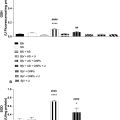
The NeoDoppler probe was attached over the infant’s fontanel and continuous recordings were made with 1 minute pauses [ ]. HITS were manually annotated as described above. To examine if HITS intensity increased at the time of median sternotomy, the HITS detected in the period from five minutes before ( t = -5) to five minutes after ( t = 5) sternotomy ( t = 0) were compared to similar ten-minute windows preceding (from t = -15 to t = -5) and following (from t = 5 to t = 15). First, HITS intensity between periods were compared using the Krusk–Wallis test and Dunn’s test with Benjamini–Hochberg correction for pairwise multiple comparisons. Second, linear mixed models were used to take interindividual differences into account by including participant as random intercept. p values were estimated with Satterthwaite’s method using the lmerTest package for R [ ].
Ethics
Parts 1 and 2 were approved by the Norwegian Animal Research Authority (FOTS ID 19803), and the studies adhered to the Norwegian Laboratory Animal Regulations and the EU directive 2010/63/EU. Part 3 was approved by the Regional Committee for Medical and Health Research Ethics, REC Central (Reference 2017/314), the Norwegian Directorate of Health (Reference 17/15181-11) and The Norwegian Medicines Agency (Reference 19/05458), and informed written consent was obtained from the parents of all participants.
Results
Part 1: HITS monitoring during femoral nailing in a porcine model
Experimental characteristics
Bilateral reamed intramedullary nailing of the femur was performed on 12 pigs (median weight 29.5 kg, 11 males, Table S1 ) as reported by Kristiansen et al. [ ]. Mean ± SD experimental duration from start of surgery to sacrifice was 275 ± 18 minutes ( Fig. 3 ).

Cerebral fat embolism from reamed intramedullary nailing of the femur
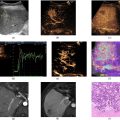
Cerebral bone marrow emboli were identified in brain biopsies from 11 of 12 animals and systemic fat emboli were demonstrated in biopsies from lungs and heart in all animals as previously reported [ ]. Brain tissues from the two animals with most HITS were restained. In total, 700 emboli were found across eight brain tissue sections ( Fig. 4 ), with median and maximum areas 11 µm 2 and 795 µm, 2 respectively. Only 11% of the emboli were larger than an erythrocyte ( Fig. 4 D). The emboli were primarily located in or close to the meninges but could also be found in the parenchyma. In addition, bone fragments were found in two sections ( Fig. 4 C).

Related posts:
 Strain Patterns With Ultrasound for Assessment of Abdominal Aortic Aneurysm Vessel Wall Biomechanics
 Evaluation of Renal Masses Using Contrast-Enhanced Ultrasound with Sonovue and Sonazoid
 A Bioengineered Cathepsin B-sensitive Gas Vesicle Nanosystem That Responds With Increased Gray-level Intensity of Ultrasound Biomicroscopic Images
 Automated Approach for Enhancing Fetal Head Station Assessment in Labor with Transperineal Ultrasound
 Classification of Neoadjuvant Therapy Response in Patients With Colorectal Liver Metastases Using Contrast-Enhanced Ultrasound—With Histological Pathology as the Gold Standard
 Ultrasound and Gold Nanoparticles Improve Tissue Repair for Muscle Injury Caused by Snake Venom
Stay updated, free articles. Join our Telegram channel

Full access? Get Clinical Tree


