Peripheral arterial ultrasound is an important technique to evaluate vascular disease. Protocols include grayscale, color Doppler and spectral Doppler. Arterial duplex is used to evaluate patients with claudication to characterize atherosclerotic disease. Stenoses have characteristic findings which can help profile lesions accurately. Other nonatherosclerotic obstructive processes are also evaluable by duplex Doppler. Traumatic injuries can also be studied in the stable patient. Attention to protocols helps ensure optimal results.
Key points
- •
Peripheral arterial duplex ultrasound with grayscale, color and spectral Doppler can define most extremity stenosis and is helpful for evaluating traumatic lesions.
- •
Criteria for stenosis differ between native arteries and graft and post intervention examinations.
- •
Deep arteries, calcified arteries and small calf arteries are more difficult to evaluate with ultrasound.
Peripheral artery
Noninvasive peripheral artery evaluation began as non-imaging physiologic investigations, for example, ankle-brachial index (ABI), to measure disease. Duplex Doppler ultrasound (US) using grayscale, color and spectral Doppler ultrasound directly localizes and measures disease. ABIs are complementary. Spectral Doppler is quantitative while color Doppler localizes the level of disease more efficiently and with confidence.
Most patients evaluated by duplex Doppler have chronic limb ischemia. Acute ischemia is generally evaluated in a more emergent fashion typically by CT or MR angiography rather than US. Arterial duplex has expanded to other processes including traumatic injuries, functional stenotic disease (eg, popliteal entrapment), arteritis, and mappings prior to interventions. ,
Anatomy
The aorta and external iliac arteries are the major inflow arteries. The internal iliac artery is an important collateral pathway from the pelvis to the limb. The common femoral artery originates at the inguinal ligament and after a few centimeters bifurcates into the superficial femoral and the deep femoral artery. The superficial femoral artery travels down the thigh and exits Hunter’s canal to become the popliteal artery. The popliteal artery terminates at the junction of the anterior tibial artery and tibioperoneal trunk. The trunk bifurcates into the posterior tibial and peroneal arteries. The posterior tibial artery, located at the ankle behind the medial malleolus and the dorsalis pedis artery (derived from the anterior tibial artery) located on the dorsum of the foot are arteries palpated during physical examination.
The left and right subclavian arteries are usually the first vessels studied during an upper extremity arterial examination. Portions of the arteries are not evaluable due to their relationship to the clavicle. The subclavian artery becomes the axillary artery and then the brachial artery, typically around the antecubital fossa. The bifurcation of the brachial artery forms the ulnar and radial arteries. The arteries at the wrist form several arches before supplying the digital arteries. The radial artery is often chosen for catheterization or even resection (during, for instance, free flap formation), since most patients are not radial artery dependent.
Clinical Overview of Atherosclerosis
Lower extremity atherosclerotic diseases are roughly classified into inflow (aorto-iliac), femoral-popliteal, and/or outflow (calf) disease.
In the earliest stages, atherosclerotic plaques are asymptomatic. As plaque grows, the lumen narrows, which produces stenoses, typically at multiple sites. With progression, symptoms of claudication appear during activity, and eventually progress from mild to disabling pain. In severe cases, resting pain or ischemic changes can result.
Multiple sites of stenosis may be present in a single vessel or at multiple vessels. Progressive stenosis or plaque rupture can lead to complete occlusion. As atherosclerotic disease advances, collateral flow may reconstitute flow, and may mitigate the course of the disease.
Overview of Peripheral Arterial Spectral Waveforms
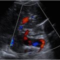
A recent consensus paper analyzed arterial and venous waveforms. Arterial phases are simplified: If the direction of the outer edge crosses the zero baseline and reverses, it is a multiphasic waveform ( Fig. 1 ). If it does not cross the baseline the waveform is monophasic ( Fig. 2 ).


The outline (envelope) of the normal multiphasic peripheral artery spectral waveform is a rapid systolic upstroke (time to peak) and sharp systolic downstroke which crosses the baseline (see Fig. 1 ). The reversal is typically brief followed by a return to baseline. This pattern may be followed by oscillating forward and backward phases. Formerly, describing waveforms as biphasic, triphasic, and so on were used; however, now these waveforms are simply lumped together as multiphasic.
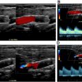
The interior of the waveform (window) under the outside envelope (representing slower velocities) is also evaluated , (see Fig. 1 ). In a normal straight artery with laminar flow, most of the flow in the center of the artery is traveling near the fastest velocity (see Fig. 2 ). For that reason, the waveform is brightest at the edge, with minimal lower velocities or no flow represented as darker to absent grays. In other arteries with parabolic flow, blood velocity gradually diminishes from the center to the edge and creates a more uniform filling in of the waveform’s interior. “Fill in” is termed spectral broadening . Spectral broadening also occurs in normal vessels as blood within different regions of the lumen flows at varying velocities, for instance, around a curve or through bifurcations ( Fig. 3 A, B ). Technical factors such as an overly large sample volume may also produce broadening. Spectral broadening is an important criterion for abnormal vessels. Irregular flow occurs beyond a stenosis: mild spectral broadening is found with milder stenosis, and turbulent flow appears with more significant stenoses. Irregular currents not aligned with the lumen are seen as multiple velocities and less definition of the edge of the spectrum and simultaneous forward and reversed flow. Vortices with higher velocity spikes may be superimposed on the slower moving blood (“picket fence” appearance).

Spectral Waveforms and Color Doppler Findings in Stenoses
Within a stenosis, narrowing initially produces little change in velocity but as the degree of stenosis increases, the velocity increases related to the drop in pressure through the stenosis. The increase in velocity is related to the degree of stenosis.
Waveforms within the stenosis are laminar and monophasic with increased systolic and diastolic velocities. In practice, systolic velocity elevation is used as the main criterion for stenosis, although some criteria might also use diastolic velocity or a pulsatility index.
Further downstream from the disturbed flow of stenosis, the flow resumes a laminar profile, but the waveform shape may change to a tardus-parvus (blunted) waveform. The tardus-parvus waveform reflects 2 significant hemodynamic changes created by a significant stenosis. The speed of the systolic upstroke may be diminished (“parvus”) and the acceleration is prolonged, taking longer than normal to reach peak (“tardus”). The diminished pressure beyond a significant stenosis reduces the difference between systolic and diastole pressure. The velocity difference in the distal waveform between systole and diastole is likewise reduced creating a less pulsatile waveform.
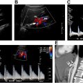
Arterial stenoses are diagnosed by a profile ( Fig. 4 A–D), with normal or low velocities before the stenosis, elevated velocities within the stenosis, and decreased velocity with post stenotic turbulence beyond the stenosis. , Relying simply on the elevated velocity is not as accurate as confirming all 3 waveforms on a duplex examination.

Mild stenoses have no or modest increased velocity with filling in of the spectral envelope or mild bidirectional disturbed flow. Significant stenoses are pressure reducing and flow limiting. These stenoses are associated with significantly elevated (especially systolic) velocities, post-stenotic turbulence beyond the immediate narrowing, and usually tardus-parvus waveforms further distally.
The waveform shape proximal to a stenosis is variable depending on the degree of stenosis, degree of collateral flow, and pressure reduction created by the stenosis. In a normal multiphasic waveform, downstream pressure creates reflected waves from downstream arterioles to produce the reversed phase. A pressure-reducing lesion produces less antegrade pressure and a significantly attenuated reflected wave, thereby reducing or eliminating the reversed components proximal to the stenosis. Vasodilatation distally may also be a factor. Therefore, the waveform before a stenosis may be monophasic with a rapid upstroke unless it has been altered by a proximal obstruction. But, if the degree of stenosis is profound or there is occlusion, the waveform proximally may be excessively pulsatile with only a short forward systolic phase (staccato, water hammer waveform) indicating very high resistance beyond the sample site.
Color Doppler is best considered a pathfinder and shows narrowing at areas of plaque formation (see Fig. 4 B). The color is not equivalent to an angiogram since the color represents a rough outline of the lumen. Factors such as gain, phase of the cardiac cycle, and low volume may affect the color diameter and intraluminal features. 2 color Doppler criteria are used for stenoses: the size of the color lumen and the change in velocity in areas of narrowing. Elevated velocity in areas of narrowing change the color (appreciated by the color scale next to the image).
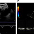
Aliasing is an artifact that can help identify high velocity flow. Aliasing occurs when the velocity which is being measured exceeds the sampling rate, so that direction and velocity are misrepresented ( Fig. 5 A, B ). Color aliasing has 2 appearances: (1) a series of layers of colors in the proper direction next to color layers encoded in the wrong direction (“onion skin” appearance) or (2) correct and incorrect direction pixels mixed (“mosaic” pattern). Aliasing is also seen in spectral Doppler where the waveform wraps around top of the correct direction and reappears at the bottom in the wrong direction. If the artifact is large enough, the wrap around even returns to the correct direction (albeit with the incorrect velocity). Spectral multiple aliasing is the analog of the mosaic pattern, but aliasing is more common in color Doppler since the sampling rate for color is far slower than spectral Doppler.

Protocols
Lower extremity native arteries
Arteries are scanned with different transducers based on the depth of the artery from the skin. , In general, the highest frequency is used, and linear transducers are favored, since this configuration aligns with the vessel wall. Curved array transducers are generally reserved for deep pelvic vessels and where body habitus is adverse. The femoral vessels in the thigh are generally scanned from an anterior position with the leg rotated outward. A posterior approach, sometimes in the decubitus position, is generally needed for popliteal and posterior calf artery evaluation. The anterior tibial artery is small and usually the most difficult calf artery to visualize, optimally imaged from the anterolateral position, although its origin may be seen from a posterior view.
The native arterial duplex mapping protocol consists of mapping from the common femoral to the popliteal arteries ( Table 1 ). All accessible portions of the arteries are evaluated since plaque and stenosis, although favoring some locations, may occur anywhere along the artery. In a patient without stenosis, representative segments are documented at selected sites.
Related posts:

Stay updated, free articles. Join our Telegram channel

Full access? Get Clinical Tree


