Peritoneal Metastases
R. Brooke Jeffrey, MD
Key Facts
Terminology
Peritoneal carcinomatosis, peritoneal implants, omental masses (“caking”)
Metastatic disease to omentum, peritoneal surface, peritoneal ligaments, &/or mesentery
Imaging
Best diagnostic clue: Omental caking, soft tissue implants on peritoneal surface
CT findings
Ascites, nodular peritoneal thickening/enhancement, hypovascular omental caking
Spiculated mesentery
Evidence of bowel obstruction with delineation of transition zone from dilated to nondilated bowel
Best imaging tool: CECT or MR
Protocol advice: Oral & IV contrast (CECT), gadolinium-enhanced GRE or T1 sequence (MR)
Top Differential Diagnoses
TB peritonitis
Papillary serous carcinoma of peritoneum
Peritoneal mesothelioma
Pseudomyxoma peritonei
Pathology
Peritoneal metastases indicate stage IV disease
Clinical Issues
Abdominal distension and pain, weight loss, ± ascites
M < F in secondary ovarian carcinoma
Poor prognosis in general
Diagnostic Checklist
TB peritonitis
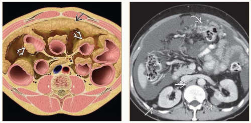 (Left) Axial anatomic rendering of peritoneal metastases. Note the anterior omental cake  and serosal implants and serosal implants  . (Right) Axial CECT shows a massive, malignant ascites due to peritoneal metastases . (Right) Axial CECT shows a massive, malignant ascites due to peritoneal metastases  . Note the metastases to the abdominal wall, lymph nodes, and perirenal space bilaterally . Note the metastases to the abdominal wall, lymph nodes, and perirenal space bilaterally  . . |
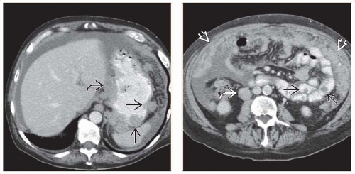 (Left) Axial CECT shows classic widespread metastases from melanoma into the gastric wall  and lymph nodes and lymph nodes  . (Right) Axial CECT in the same patient demonstrates the spread of metastases to the small bowel . (Right) Axial CECT in the same patient demonstrates the spread of metastases to the small bowel  , lymph nodes , lymph nodes  , and omentum. Both nodular and diffuse metastases are seen , and omentum. Both nodular and diffuse metastases are seen  . . |
TERMINOLOGY
Synonyms
Peritoneal carcinomatosis, peritoneal implants, omental masses (“caking”)
Definitions
Metastatic disease to omentum, peritoneal surface, peritoneal ligaments, &/or mesentery
IMAGING
General Features
Best diagnostic clue
Omental caking, soft tissue implants on peritoneal surface
Cystic peritoneal masses with ovarian carcinoma on peritoneal surfaces
Ascites, mesenteric stranding, bowel obstruction
Location
Peritoneum, mesentery, peritoneal ligaments
Size
Variable; 5 mm nodules to large confluent omental caking
Morphology
Nodular, plaque-like, or large omental mass
Radiographic Findings
Radiography
Plain film findings of significant ascites
Medial displacement of cecum in 90% of patients
Pelvic “dog’s ear” in 90% of patients
Medial displacement of lateral liver edge (Hellmer sign) in 80% of patients
Bulging of flanks, central displacement of bowel loops, indistinct psoas margin
Related posts:

Stay updated, free articles. Join our Telegram channel

Full access? Get Clinical Tree


