KEY FACTS
Imaging
- •
Exclusively in male fetuses
- •
Bladder often grossly dilated, filling entire abdomen and increasing abdominal circumference
- •
Distended bladder “funnels” into urethra
- ○
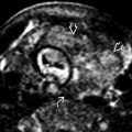
Keyhole sign from dilated posterior urethra
- ○
- •
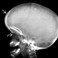
Small, bell-shaped chest
- •
Hydronephrosis/hydroureter
- •
Rupture → urinoma or urinary ascites
- •
Oligohydramnios or anhydramnios in severe cases
- •
Kidneys may show signs of obstructive renal dysplasia
Top Differential Diagnoses
- •
Cloacal malformation seen exclusively in females
- ○
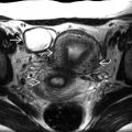
Dilated, fluid-filled vagina can mimic bladder
- ○
- •
Prune-belly syndrome
Clinical Issues
- •
Most common cause of fetal lower urinary tract obstruction
- •
Variable degree of fetal renal damage, directly affects long-term outcomes and survival (overall mortality: 25-50%)
- •
May tap fetal bladder and assess urine electrolytes
- ○
Intervention (e.g., vesicoamniotic shunt) may be considered in cases with good prognostic markers
- ○
Scanning Tips
- •
If bladder appears large, check several times during exam to see if it decompresses
- ○
Likely normal if otherwise normal urinary tract and normal amniotic fluid
- ○
- •
Determine sex
- ○
May be difficult in setting of oligohydramnios
- ○
If female, what appears to be a dilated bladder may actually be vagina
- ○
- •
Obtain focused images of bladder base to evaluate for dilation of posterior urethra (keyhole)
- •
Evaluate kidneys for signs of renal parenchymal damage
- ○
Increased echogenicity, small cortical cysts, atrophy
- ○
 . The valve forms a thin membrane
  , blocking antegrade flow of urine and creating a lower urinary tract obstruction (LUTO).
 , with severe oligohydramnios and a small, bell-shaped chest
  . The inset shows dilatation of the posterior urethra
  , the keyhole sign. This is the “key” to the diagnosis and differentiates posterior urethral valves from other causes of LUTO.
Stay updated, free articles. Join our Telegram channel

Full access? Get Clinical Tree


