Premotor Cortex and Supplementary Motor Area (Area 6)
Jared A. Nielsen, PhD
Key Facts
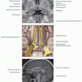
Location and Boundaries
Caudal portions of inferior and middle frontal gyri and inferior frontal sulcus
Caudal portions of middle and lateral superior frontal gyri and superior frontal sulcus
Caudal and medial portion of superior frontal gyrus
Portion of medial superior frontal gyrus
Function
Supplementary motor area (SMA) selects actions initiated from internal cues
Premotor cortex selects actions initiated from external cues
Motor planning
Coordinate action sequence
Voluntary eye movements
Acquisition of motor skills
Timing of action execution
Motor flexibility
Inhibit action
Change action plan
Initiate new action
Structural Connections
Primary sensorimotor cortex (areas 1, 2, 3, and 4)
Superior parietal cortex (areas 5, 7)
Supramarginal gyrus (area 40)
Corticospinal and corticobulbar tracts
Functional Connections
Primary sensorimotor cortex (areas 1, 2, 3, and 4)
Intraparietal sulcus (areas 5, 7)
Thalamus
Putamen
Globus pallidus
Cerebellum
NeuroSynth keywords: Saccade, eye, shift, movement, spatial, execution, load, position, attention
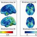
 (Left) Coactivation map of Brodmann area 6 shows brain regions that reliably activate with the centroid of voxels lying within area 6 in over 4,000 studies from the NeuroSynth database. Image is the average of left and right coactivation maps. |
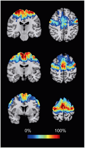 (Right) Cytoarchitectonic map of premotor cortex and SMA represents quantitative probabilistic map derived from postmortem human brains and is specific to cellular properties unique to area 6 (data source: SPM Anatomy toolbox). |
LOCATION AND BOUNDARIES
Location
Dorsal premotor cortex
Caudal portions of inferior and middle frontal gyri and inferior frontal sulcus
Includes frontal eye fields at confluence of superior frontal and precentral sulci
Rostral to hand area of primary motor cortex (area 4) and caudal to superior prefrontal cortex (area 8)
Ventral premotor cortex
Caudal portions of middle and lateral superior frontal gyri and superior frontal sulcus
Rostral to face area of primary motor cortex (area 4) and caudal to Broca area (in particular area 44)
Supplementary motor area
Caudal and medial portion of superior frontal gyrus
Rostral to leg area of primary motor cortex (area 4) and caudal to presupplementary motor area
Presupplementary motor area
Portion of medial superior frontal gyrus
Rostral to supplementary motor area and caudal to superior prefrontal cortex (area 8)
Boundaries
Caudal: Precentral gyrus
Medial and ventral: Cingulate sulcus
Lateral and ventral: Lateral sulcus
Rostral: Ventral portions of inferior (area 44), middle (area 9), and superior frontal (area 8) gyriRelated posts:

Stay updated, free articles. Join our Telegram channel

Full access? Get Clinical Tree