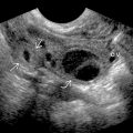
KEY FACTS
Imaging
- •
Grayscale US
- ○
Prostate carcinoma (PCa) can appear as hypoechoic (60-70%), isoechoic/invisible (30-40%), rarely hyperechoic ± asymmetric capsular bulging or irregularity
- ○
- •
Color Doppler US
- ○
PCa may be hypervascular; however, absence does not exclude cancer, and other benign entities (e.g., prostatitis) may also be hypervascular
- ○
- •
Transrectal US (TRUS) is imaging of choice to guide biopsy in evaluation for PCa but performs poorly in cancer detection and staging
- •
Calculate prostate volume using largest cross-sectional image in transverse and mid sagittal planes: Transverse x AP x long x 0.52
- •
MR imaging
- ○
Most sensitive imaging technique for PCa diagnosis and staging
- ○
Peripheral zone (PZ): T2 dark lesion with restricted diffusion; transition zone (TZ): Erased charcoal sign on T2WI
- ○
Targeted MR-guided and MR/US fusion-guided biopsy is promising for increasing detection of high-risk prostate cancer while reducing detection of low-risk cancer compared with standard biopsy
- ○
Pathology
- •
Most common noncutaneous malignancy in western world, 2nd most common cause of cancer deaths among men
- •
95% of tumors are acinar adenocarcinoma
- •
Staging based on tumor-node-metastasis staging, prostate specific antigen at time of diagnosis, and Gleason score
- •
Location of prostate cancer: PZ: 70-80%, TZ: 20%, central zone: 1-5%
Clinical Issues
- •
Management is complex due to difficulty in accurate staging and in predicting speed of disease progression
- •
Despite having higher volumes and prostate-specific antigen values at diagnosis, TZ cancers are less likely to be associated with seminal vesicle, extraprostatic and lymphovascular invasion
Scanning Tips
- •
Although classically PCa is hypoechoic on grayscale US, > 30-40% PCa may be invisible on TRUS
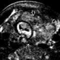
 , rectal wall
  , and symphysis pubis
  .
.

 with capsular bulging and nodularity extending into the adjacent fat
  and left neurovascular bundle
  . A smaller hypoechoic lesion is seen in the right PZ
  .
.
 in the left PZ lesion. The right PZ lesion is mildly hypervascular
  . Prostatectomy revealed multifocal PCa, including Gleason 5 + 5 adenocarcinoma with left extraprostatic extension and lymphovascular and perineural invasion.
 . About 70% of prostate cancers occur in the peripheral zone.
 around the mass. The increased vascularity of the left lobe may be due to its dependent position.


