Protocols for fMRI Examination
Jeffrey S. Anderson, MD, PhD
Key Facts
Concepts
Anatomic scan (MPRAGE [T1] &/or T2): 3-6 minutes per scan
Field map (for optional distortion correction): 3-5 minutes
Diffusion tensor imaging: 4-10 minutes, depending on number of directions, averages used
BOLD task sequences: 2-8 tasks of 4-6 minutes each
BOLD resting state: 5-50 minutes of imaging, may be broken into blocks
Total scan time as clinically required and tolerated by patient: Usually 30-60 minutes
Applications
Use T2 or 3D FLAIR anatomic image for tumors best seen on fluid-sensitive sequences
If contrast is desired, acquire BOLD images first, then MPRAGE or stereotactic 3D T1 sequence post contrast
Patients often have difficulty holding still for longer sequences; target 4-7 minutes for each sequence
Repeat key task sequences for improved signal to noise ratio
Task runs can be used for resting-state analyses, but there will be systematic changes in functional connectivity if comparing to normative data acquired during resting state
Use same slice locations and thickness for field map and BOLD acquisitions
Parallel imaging is helpful for most pulse sequences if available, particularly at 3T
Although some BOLD tasks activate only limited brain slices, whole-brain coverage is advised as coregistration to anatomic image is problematic for partial-brain acquisitions
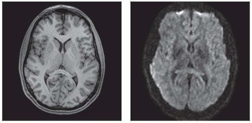 (Left) MPRAGE allows excellent gray matter/white matter contrast to facilitate normalization to a template brain space, segmentation of gray matter and white matter, and volumetric or gray matter density analysis. (Right) BOLD shows relatively poor contrast of anatomic structures and low resolution (64 × 64 matrix). T2* changes over time are related to functional brain activation. |
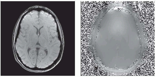 (Left) Field map (magnitude image) combining phase and magnitude images allows construction of an image that can be used to perform distortion correction. Distortion in areas of high magnetic susceptibility can create misregistration between BOLD and MPRAGE images. (Right) Field map (phase image) is shown. Phase and magnitude GRE field map images require acquisition at 2 different echo times (TE). This will generate 3 series: 2 magnitude, and 1 phase series that can be used to generate a voxel displacement map. |
Related posts:
Stay updated, free articles. Join our Telegram channel

Full access? Get Clinical Tree


