Radiographic Signs of ACL Tear
Ryan E. Embertson
Daniel B. Nissman
FINDINGS
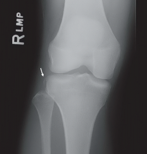
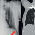
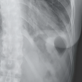
Lateral radiograph of the right knee (Fig. 8A) reveals a moderate- to large-sized effusion and a deep lateral sulcus terminalis (arrow). Anteroposterior (AP) radiograph of the right knee in a different patient (Fig. 8B) reveals a vertically oriented curvilinear bone fragment (arrow) adjacent to the lateral aspect of the lateral tibial plateau.
DIFFERENTIAL DIAGNOSIS
First case: normal variant, impaction fracture, impaction fracture with ligament injury. Second case: ligament avulsion fracture, fracture caused by direct trauma, osteophyte, accessory ossicle, heterotopic ossification.
DIAGNOSIS
First case: deep lateral femoral condyle sulcus terminalis. Second case: Segond fracture.
DISCUSSION
One of the more common injuries to the knee, especially in sports such as football, soccer, and skiing, is a tear of the anterior cruciate ligament (ACL). The ACL is an intercondylar ligament that attaches proximally at the medial posterior aspect of the lateral femoral condyle and distally at the medial aspect of the intercondylar eminence of the anterior tibia; its function is to prevent anterior translation of the tibia as well as to prevent hyperextension of the knee. Common mechanisms of ACL tear include valgus stress on the knee with the leg in relative extension, and a pivot shift. Pivot shift injury occurs when there is a valgus force applied to the flexed knee in combination with an external rotation of the tibia or an internal rotation of the femur, which occurs when there is rapid deceleration with simultaneous direction change. The diagnosis of a deficient ACL is often missed on clinical and even radiographic grounds; in one study, the correct diagnosis was made in only 9.8% of patients on initial presentation.1 The cost of missing the diagnosis is the potential for early onset osteoarthrosis resulting from loss of the stabilizing effect of the ACL.
Stay updated, free articles. Join our Telegram channel

Full access? Get Clinical Tree


